Review
. 2022 Sep 4;23(17):10135.
doi: 10.3390/ijms231710135.

Factors and Pathways Modulating Endothelial Cell Senescence in Vascular Aging

Affiliations
Review

Factors and Pathways Modulating Endothelial Cell Senescence in Vascular Aging

Hyun Jung Hwang et al. Int J Mol Sci. .

Abstract

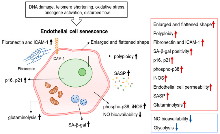
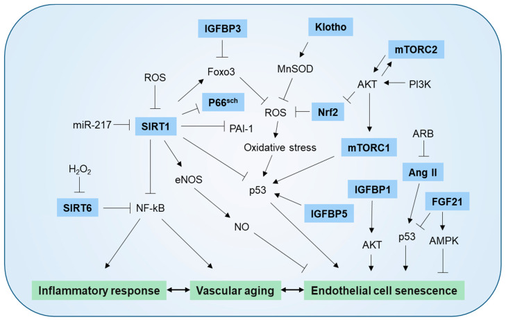
Aging causes a progressive decline in the structure and function of organs. With advancing age, an accumulation of senescent endothelial cells (ECs) contributes to the risk of developing vascular dysfunction and cardiovascular diseases, including hypertension, diabetes, atherosclerosis, and neurodegeneration. Senescent ECs undergo phenotypic changes that alter the pattern of expressed proteins, as well as their morphologies and functions, and have been linked to vascular impairments, such as aortic stiffness, enhanced inflammation, and dysregulated vascular tone. Numerous molecules and pathways, including sirtuins, Klotho, RAAS, IGFBP, NRF2, and mTOR, have been implicated in promoting EC senescence. This review summarizes the molecular players and signaling pathways driving EC senescence and identifies targets with possible therapeutic value in age-related vascular diseases.

Keywords: age-related vascular disease; cellular senescence; endothelial cell; molecular player; putative target; signaling pathway.


Conflict of interest statement

The authors declare no conflict of interest.

Figures

Figure 1
Figure 1
Characteristics of senescent endothelial cells. Various stimuli including DNA damage, telomere shortening, disturbed flow induce endothelial cell (EC) senescence. EC senescence is a state of indefinite cell cycle arrest accompanied by multiple biochemical and metabolic changes. Senescent cells generally become enlarged and flattened and display increased polyploidy, SA-β-gal activity, permeability, phosphorylation of p38, and expression of p16/p21. In addition, senescent ECs display elevated fibronectin, ICAM-1, and iNOS and reduced of NO bioavailability. Senescent ECs also produce a complex and unique SASP that includes secretion of IL-8, IL-6, PAI-1, MCP-1, IGFBP-5, and CXCL11. The metabolic alterations of senescent ECs include reduced glycolysis and increased glutaminolysis.
Figure 2
Figure 2
Molecular mechanism mediating endothelial cell senescence and vascular aging. SIRT1 protects against endothelial dysfunction by deacetylating eNOS and increasing endothelial NO bioavailability. SIRT1 also protects vascular inflammation by suppressing NF-κB signaling and exerts anti-oxidant action by increasing Foxo and down-regulating p66shc. Under conditions of age-related overexpression, miR-217 suppresses SIRT1 production. Klotho increases MnSOD activity and NO availability by negatively downregulating IGF-1 signaling. H2O2 induces endothelial cell (EC) senescence via downregulation of SIRT6 levels. SIRT6 deficiency raises the expression of endothelial proinflammatory cytokines and increases NF-κB transcriptional activity. Increased oxidative stress due to NRF2 deficiency promotes EC senescence. mTORC1 increases EC senescence through the PI3K/AKT pathway and mTORC2 induces EC senescence by suppressing NRF2 production via the AKT/GSK-3β/CEBPα signaling pathway. IGFBP1 and IGFBP5 induce senescence in HUVECs through AKT or p53-dependent mechanism. IGFBP3 promotes EC senescence by inhibiting the SIRT1-Foxo axis, ARB inhibits Ang II-induced EC senescence, and FGF21 represses EC senescence via inhibiting p53 signaling pathway in an AMPK-dependent manner.

References

    1. Muñoz-Espín D., Serrano M. Cellular senescence: From physiology to pathology. Nat. Rev. Mol. Cell Biol. 2014;15:482–496. doi: 10.1038/nrm3823. - DOI - PubMed
    1. Chicas A., Wang X., Zhang C., McCurrach M., Zhao Z., Mert O., Dickins R.A., Narita M., Zhang M., Lowe S.W. Dissecting the unique role of the retinoblastoma tumor suppressor during cellular senescence. Cancer Cell. 2010;17:376–387. doi: 10.1016/j.ccr.2010.01.023. - DOI - PMC - PubMed
    1. Muñoz-Espín D., Cañamero M., Maraver A., Gómez-López G., Contreras J., Murillo-Cuesta S., Rodríguez-Baeza A., Varela-Nieto I., Ruberte J., Collado M., et al. Programmed cell senescence during mammalian embryonic development. Cell. 2013;155:1104–1118. doi: 10.1016/j.cell.2013.10.019. - DOI - PubMed
    1. Demaria M., Ohtani N., Youssef S.A., Rodier F., Toussaint W., Mitchell J.R., Laberge R.M., Vijg J., Van Steeg H., Dollé M.E., et al. An essential role for senescent cells in optimal wound healing through secretion of PDGF-AA. Dev. Cell. 2014;31:722–733. doi: 10.1016/j.devcel.2014.11.012. - DOI - PMC - PubMed
    1. Feng T., Meng J., Kou S., Jiang Z., Huang X., Lu Z., Zhao H., Lau L.F., Zhou B., Zhang H. CCN1-Induced Cellular Senescence Promotes Heart Regeneration. Circulation. 2019;139:2495–2498. doi: 10.1161/CIRCULATIONAHA.119.039530. - DOI - PubMed