. 2022 Sep 4;23(17):10146.
doi: 10.3390/ijms231710146.

Dapagliflozin Mitigates Doxorubicin-Caused Myocardium Damage by Regulating AKT-Mediated Oxidative Stress, Cardiac Remodeling, and Inflammation

Affiliations

Dapagliflozin Mitigates Doxorubicin-Caused Myocardium Damage by Regulating AKT-Mediated Oxidative Stress, Cardiac Remodeling, and Inflammation

Pei-Ling Hsieh et al. Int J Mol Sci. .

Abstract

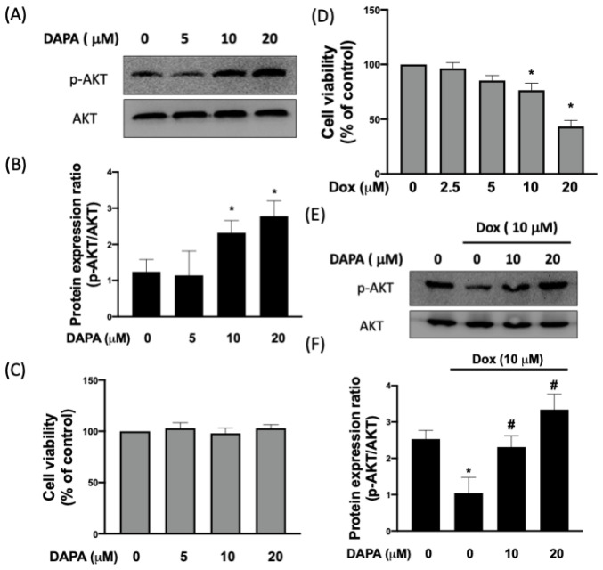
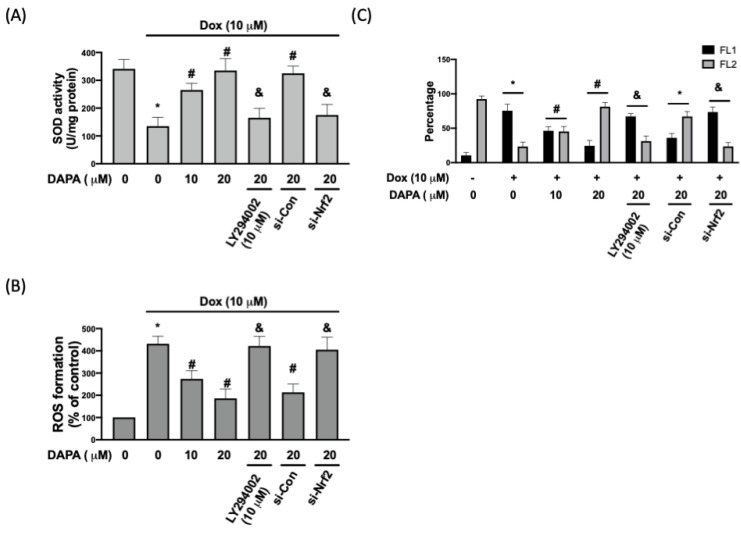
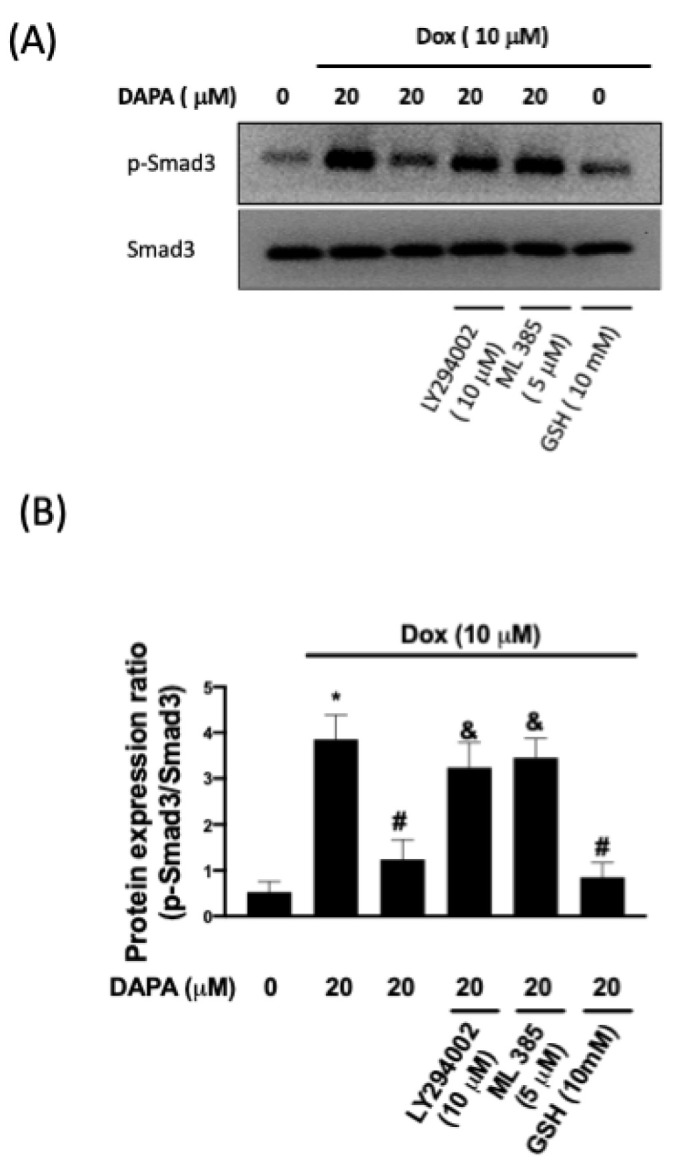
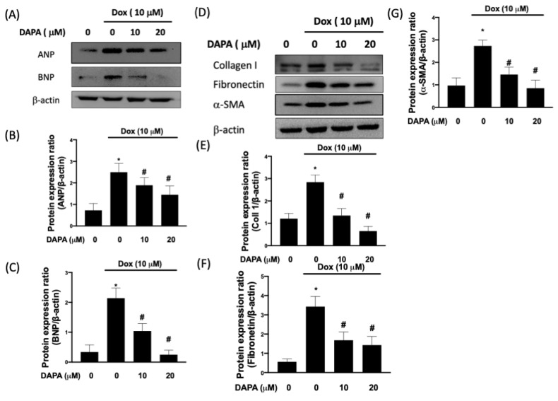
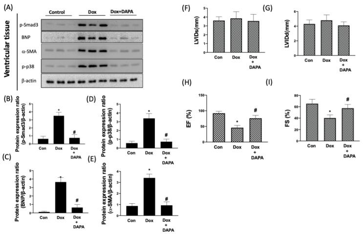
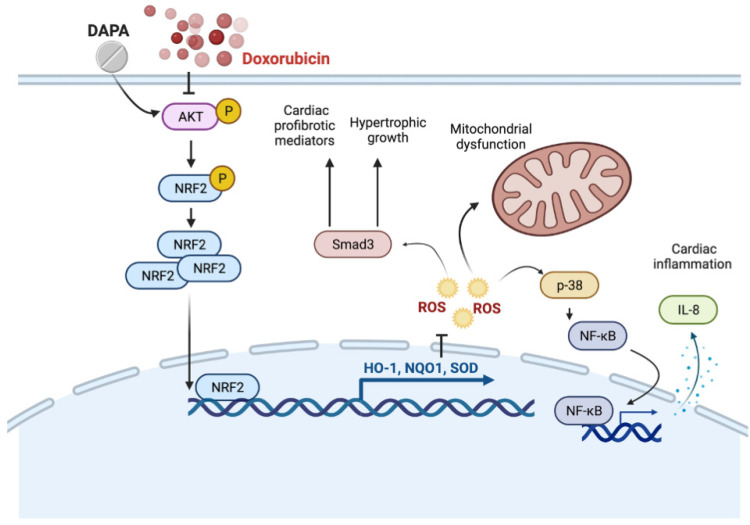
Doxorubicin (Dox) is a commonly used anthracycline chemotherapy with a side effect of cardiotoxicity, which may increase the risk of heart failure for cancer patients. Although various studies have demonstrated the cardioprotective property of dapagliflozin (DAPA), a sodium-glucose cotransporter 2 inhibitor, the detailed mechanism underlying its effect on Dox-induced cardiomyopathy is still limited. In this study, we showed that DAPA induced the activation of AKT/PI3K signaling in cardiac myoblast H9c2 cells following Dox treatment, leading to the upregulation of antioxidant HO-1, NQO1, and SOD, as well as an improved mitochondrial dysfunction via Nrf2. In addition, the reduced oxidative stress resulted in the downregulation of hypertrophy (ANP and BNP) and fibrosis (phospho-Smad3, collagen I, fibronectin, and α-SMA) markers. Furthermore, the inflammatory IL-8 concentration was inhibited after DAPA, possibly through PI3K/AKT/Nrf2/p38/NF-κB signaling. Moreover, our results were validated in vivo, and echocardiography results suggested an improved cardiac function in DAPA-receiving rats. In summary, we demonstrated that the administration of DAPA could mitigate the Dox-elicited cardiotoxicity by reducing oxidative stress, mitochondrial dysfunction, fibrosis, hypertrophy, and inflammation via PI3K/AKT/Nrf2 signaling.

Keywords: Akt; cardiotoxicity; dapagliflozin; doxorubicin.


Conflict of interest statement

The authors declare no conflict of interest.

Figures

Figure 1
Figure 1
Treatment of DAPA restores the Dox-inhibited phosphorylation of AKT. Representative Western blot images (A) and densitometric analysis (B) of phosphorylated and total AKT levels in H9c2 cells treated with various concentrations of DAPA. Cell viability of H9c2 cells after DAPA treatment (C). The dosage-dependent manner of Dox exposure (D). Representative Western blot images (E) and densitometric analysis (F) of phosphorylated and total AKT levels in Dox-stimulated H9c2 cells following DAPA co-administration were shown. Results are shown as means ± SD of three independent experiments. Statistical analysis was performed using one-way ANOVA with a Bonferroni post hoc test. [* p < 0.05 compared to the control group; # p < 0.05 compared to the Dox-only group].
Figure 2
Figure 2
DAPA administration prevents the Dox-inhibited Nrf2 nuclear translocation and expression of HO-1 and NQO1. Western blot images (A), densitometric analysis of nuclear fraction (B), and cytosolic fraction (C) of Nrf2 in Dox-treated H9c2 cells were shown. Representative Western blot images (D), densitometric analysis of HO-1 (E) and NQO1 (F) in Dox-stimulated cells with or without DAPA treatment were revealed. Expression levels of HO-1 and NQO1 gene in Dox-treated cells with or without DAPA treatment were checked (G). LY294002 (a PI3K inhibitor) and si-Nrf2 were used in some experiments. Results are shown as means ± SD of three independent experiments. Statistical analysis was performed using one-way ANOVA with a Bonferroni post hoc test. [* p < 0.05 compared to the control group; # p < 0.05 compared to the Dox-only group; & p < 0.05 compared to Dox + DAPA group].
Figure 3
Figure 3
DAPA diminishes Dox-induced oxidative stress and mitochondrial damage. (A) SOD activity, (B) ROS formation, and (C) percentage of cells expressing JC-1 monomers (green fluorescence; FL1) and JC-1 aggregates (red fluorescence; FL2) in Dox-treated cells with or without DAPA treatment were assessed. Results are shown as means ± SD of three independent experiments. Statistical analysis was performed using one-way ANOVA with a Bonferroni post hoc test. [* p < 0.05 compared to the control group; # p < 0.05 compared to the Dox-only group; & p < 0.05 compared to Dox + DAPA group].
Figure 4
Figure 4
Phosphorylation of Smad3 in Dox-treated cells. Representative Western blot images (A) and densitometric analysis (B) of phospho-Smad3 and total Smad3 in H9c2 cells exposed to Dox stimulation with or without DAPA treatment were shown. LY294002 (a PI3K inhibitor), ML385 (a Nrf2 inhibitor), and GSH (an antioxidant) were used in some experiments. Results are shown as means ± SD of three independent experiments. Statistical analysis was performed using one-way ANOVA with a Bonferroni post hoc test. [* p < 0.05 compared to the control group; # p < 0.05 compared to the Dox-only group; & p < 0.05 compared to Dox + DAPA group].
Figure 5
Figure 5
DAPA mitigates the expression of cardiac fibrosis markers in Dox-stimulated cells. Representative Western blot images (A) and densitometric analysis of ANP (B) and BNP (C) in Dox-treated H9c2 cells with various concentrations of DAPA treatment were shown. Representative Western blot images (D), densitometric analysis collagen I (E), fibronectin (F), and α-SMA (G) in Dox-treated H9c2 cells with various concentrations of DAPA treatment were disclosed. Results are shown as means ± SD of three independent experiments. Statistical analysis was performed using one-way ANOVA with a Bonferroni post hoc test. [* p < 0.05 compared to the control group; # p < 0.05 compared to the Dox-only group].
Figure 6
Figure 6
DAPA treatment suppresses the production of Dox-induced inflammatory mediators. Representative Western blot images (A) and densitometric analysis of phospho-p38 (B) and NF-κB p65 (C) in Dox-stimulated H9c2 cells with various concentrations of DAPA treatment were shown. NF-κB p65 activation (D) and IL-8 production (E) in Dox-treated H9c2 cells with various concentrations of DAPA treatment were examined by ELISA assay. LY294002 (a PI3K inhibitor) and si-Nrf2 were used in some experiments. The finding of NF-κB p65 activation (F) and IL-8 production (G) were conducted in primary cardiomyocytes. Results are shown as means ± SD of three independent experiments. Statistical analysis was performed using one-way ANOVA with a Bonferroni post hoc test. [* p < 0.05 compared to the control group; # p < 0.05 compared to the Dox-only group; & p < 0.05 compared to Dox + DAPA group].
Figure 7
Figure 7
DAPA protects heart function in response to Dox treatment. Representative Western blot images (A) and densitometric analysis of phospho-Smad3 (B), BNP (C), α-SMA (D), and phospho-p38 (E) in control (CON) group, Dox only group, and combination treatment of Dox and DAPA (Dox + DAPA) group were shown. (F) Left ventricular internal dimension at end-diastole (LVIDd), (G) left ventricular internal dimension at end-systole (LVIDs), (H) ejection fraction (EF), and (I) fractional shortening (FS) of the heart were measured using echocardiography. Results are shown as means ± SD of three independent experiments. Statistical analysis was performed using one-way ANOVA with a Bonferroni post hoc test. [* p < 0.05 compared to the control group; # p < 0.05 compared to the Dox-only group].
Figure 8
Figure 8
Schematic diagram of the major study findings. This study presents that DAPA reduces Dox-caused myocardial damage by regulating AKT-mediated oxidative stress, cardiac remodeling, and inflammation.

References

    1. Cardinale D., Colombo A., Bacchiani G., Tedeschi I., Meroni C.A., Veglia F., Civelli M., Lamantia G., Colombo N., Curigliano G., et al. Early detection of anthracycline cardiotoxicity and improvement with heart failure therapy. Circulation. 2015;131:1981–1988. doi: 10.1161/CIRCULATIONAHA.114.013777. - DOI - PubMed
    1. Meléndez G.C., Jordan J.H., D’Agostino R.B., Jr., Vasu S., Hamilton C.A., Hundley W.G. Progressive 3-Month Increase in LV Myocardial ECV After Anthracycline-Based Chemotherapy. JACC Cardiovasc. Imaging. 2017;10:708–709. doi: 10.1016/j.jcmg.2016.06.006. - DOI - PMC - PubMed
    1. McMurray J.J.V., Solomon S.D., Inzucchi S.E., Køber L., Kosiborod M.N., Martinez F.A., Ponikowski P., Sabatine M.S., Anand I.S., Bělohlávek J., et al. Dapagliflozin in Patients with Heart Failure and Reduced Ejection Fraction. N. Engl. J. Med. 2019;381:1995–2008. doi: 10.1056/NEJMoa1911303. - DOI - PubMed
    1. Wiviott S.D., Raz I., Bonaca M.P., Mosenzon O., Kato E.T., Cahn A., Silverman M.G., Zelniker T.A., Kuder J.F., Murphy S.A., et al. Dapagliflozin and Cardiovascular Outcomes in Type 2 Diabetes. N. Engl. J. Med. 2019;380:347–357. doi: 10.1056/NEJMoa1812389. - DOI - PubMed
    1. Takemura G., Fujiwara H. Doxorubicin-induced cardiomyopathy from the cardiotoxic mechanisms to management. Prog. Cardiovasc. Dis. 2007;49:330–352. doi: 10.1016/j.pcad.2006.10.002. - DOI - PubMed

LinkOut - more resources