. 2022 Sep 5;23(17):10184.
doi: 10.3390/ijms231710184.

Systemic Beta-Hydroxybutyrate Affects BDNF and Autophagy into the Retina of Diabetic Mice

Affiliations

Systemic Beta-Hydroxybutyrate Affects BDNF and Autophagy into the Retina of Diabetic Mice

Maria Consiglia Trotta et al. Int J Mol Sci. .

Abstract

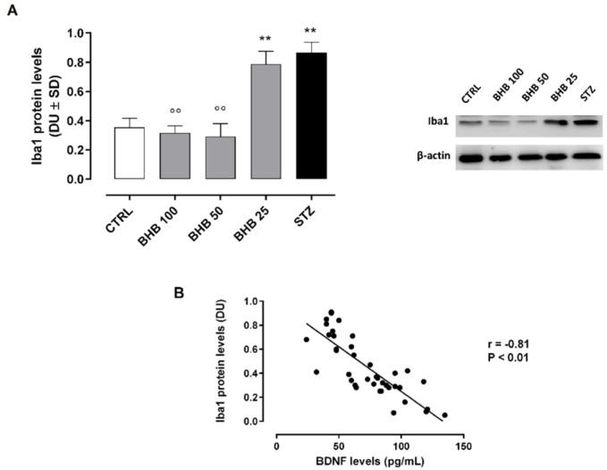
Background: Diabetic retinopathy (DR) is a neurovascular disease, characterized by a deficiency of brain-derived neurotrophic factor (BDNF), a regulator of autophagy. Beta-hydroxybutyrate (BHB), previously reported as a protective agent in DR, has been associated with BDNF promotion. Here, we investigated whether systemic BHB affects the retinal levels of BDNF and local autophagy in diabetic mice with retinopathy; Methods: C57BL/6J mice were administered with intraperitoneal (i.p.) streptozotocin (STZ) (75 mg/kg) injection to develop diabetes. After 2 weeks, they received i.p. injections of BHB (25−50−100 mg/kg) twice a week for 10 weeks. Retinal samples were collected in order to perform immunofluorescence, Western blotting, and ELISA analysis; Results: BHB 50 mg/kg and 100 mg/kg significantly improved retinal BDNF levels (p < 0.01) in diabetic mice. This improvement was negatively associated with autophagosome−lysosome formations (marked by LC3B and ATG14) and to higher levels of connexin 43 (p < 0.01), a marker of cell integrity. Moreover, BHB administration significantly reduced M1 microglial activation and autophagy (p < 0.01); Conclusions: The systemic administration of BHB in mice with DR improves the retinal levels of BDNF, with the consequent reduction of the abnormal microglial autophagy. This leads to retinal cell safety through connexin 43 restoration.

Keywords: autophagy; beta-hydroxybutyrate; brain-derived neurotrophic factor; diabetic retinopathy; hydroxycarboxylic acid receptor 2; microglia.


Conflict of interest statement

The authors declare no conflict of interest.

Figures

Figure 1
Figure 1
Retinal levels of BDNF (A), PI3K (B), LC3B (C), and strength of association between BDNF and LC3B (D) in non-diabetic mice (CTRL), diabetic mice (STZ), and diabetic mice treated with BHB 25 mg/kg (BHB 25), 50 mg/kg (BHB 50), and 100 mg/kg (BHB 100). BDNF (pg/mL) and LC3B (densitometric units, DU) levels are expressed as mean ± SD of N = 8 retinas per group. ** p < 0.01 vs. CTRL; °° p < 0.01 vs. STZ.
Figure 2
Figure 2
Retinal ATG14 levels (A) and their strength of association with BDNF (B) in non-diabetic mice (CTRL), diabetic mice (STZ), and diabetic mice treated with BHB 25 mg/kg (BHB 25), 50 mg/kg (BHB 50), and 100 mg/kg (BHB 100). ATG14 levels (ng/mL) are expressed as mean ± SD of N = 8 retinas per group. ** p < 0.01 vs. CTRL; °° p < 0.01 vs. STZ.
Figure 3
Figure 3
Retinal Cnx43 levels (A) and their strength of association with LC3B and ATG14 (B) in non-diabetic mice (CTRL), diabetic mice (STZ), and diabetic mice treated with BHB 25 mg/kg (BHB 25), 50 mg/kg (BHB 50), and 100 mg/kg (BHB 100). Cnx43 levels (ng/mL) are expressed as mean ± SD of N = 8 retinas per group. * p < 0.05 and ** p < 0.01 vs. CTRL; °° p < 0.01 vs. STZ.
Figure 4
Figure 4
Retinal Iba1 levels (A) and their strength of association with BDNF (B) in non-diabetic mice (CTRL), diabetic mice (STZ), and diabetic mice treated with BHB 25 mg/kg (BHB 25), 50 mg/kg (BHB 50), and 100 mg/kg (BHB 100). Iba1 levels (densitometric units, DU) are expressed as mean ± SD of N = 8 retinas per group. ** p < 0.01 vs. CTRL; °° p < 0.01 vs. STZ.
Figure 5
Figure 5
Detection of LC3b immunolabeling in Iba1-positive microglia (A) and relative graph (B) in non-diabetic mice (CTRL), diabetic mice (STZ), and diabetic mice treated with BHB 25 mg/kg (BHB 25), 50 mg/kg (BHB 50), and 100 mg/kg (BHB 100). (A) Sagittal retina sections of visible retinal pigment epithelium (RPE) and outer nuclear layer (ONL), showing the higher co-localization (arrow) of Iba1 (red) and LC3B (green) in the ONL of STZ and BHB 25 groups. Magnification ×63. (B) Graph expressing the percentage (%) of LC3B-positive Iba1 cells on total Iba1 cells counted. The results are reported as mean ± SD of N = 8 retinas per group. ** p < 0.01 vs. CTRL; °° p < 0.01 vs. STZ.
Figure 5
Figure 5
Detection of LC3b immunolabeling in Iba1-positive microglia (A) and relative graph (B) in non-diabetic mice (CTRL), diabetic mice (STZ), and diabetic mice treated with BHB 25 mg/kg (BHB 25), 50 mg/kg (BHB 50), and 100 mg/kg (BHB 100). (A) Sagittal retina sections of visible retinal pigment epithelium (RPE) and outer nuclear layer (ONL), showing the higher co-localization (arrow) of Iba1 (red) and LC3B (green) in the ONL of STZ and BHB 25 groups. Magnification ×63. (B) Graph expressing the percentage (%) of LC3B-positive Iba1 cells on total Iba1 cells counted. The results are reported as mean ± SD of N = 8 retinas per group. ** p < 0.01 vs. CTRL; °° p < 0.01 vs. STZ.
Figure 6
Figure 6
Retinal levels of BDNF (A) and Cnx43 (B) in non-diabetic mice (CTRL), diabetic mice treated with PTX (1 µg/100 µL) (PTX) and diabetic mice treated with both PTX and BHB 25 mg/kg (PTX-BHB 25), 50 mg/kg (PTX-BHB 50), and 100 mg/kg (PTX-BHB 100). BDNF (pg/mL) and Cnx43 (ng/mL) levels are expressed as mean ± SD of N = 8 retinas per group. ** p < 0.01 vs. CTRL.

References

    1. Altmann C., Schmidt M.H.H. The Role of Microglia in Diabetic Retinopathy: Inflammation, Microvasculature Defects and Neurodegeneration. Int. J. Mol. Sci. 2018;19:110. doi: 10.3390/ijms19010110. - DOI - PMC - PubMed
    1. Afarid M., Namvar E., Sanie-Jahromi F. Diabetic Retinopathy and BDNF: A Review on Its Molecular Basis and Clinical Applications. J. Ophthalmol. 2020;2020:1–7. doi: 10.1155/2020/1602739. - DOI - PMC - PubMed
    1. Seki M., Tanaka T., Nawa H., Usui T., Fukuchi T., Ikeda K., Abe H., Takei N. Involvement of Brain-Derived Neurotrophic Factor in Early Retinal Neuropathy of Streptozotocin-Induced Diabetes in Rats. Diabetes. 2004;53:2412–2419. doi: 10.2337/diabetes.53.9.2412. - DOI - PubMed
    1. Taşlipinar Uzel A.G., UĞurlu N., Toklu Y., Çİçek M., Boral B., Şener B., ÇaĞil N. Relationship between stages of diabetic retinopathy and levels of brain-derived neurotrophic factor in aqueous humor and serum. Retina. 2020;40:121–125. doi: 10.1097/IAE.0000000000002355. - DOI - PubMed
    1. Adornetto A., Gesualdo C., Laganà M.L., Trotta M.C., Rossi S., Russo R. Autophagy: A Novel Pharmacological Target in Diabetic Retinopathy. Front. Pharmacol. 2021;12:695267. doi: 10.3389/fphar.2021.695267. - DOI - PMC - PubMed