. 2022 Aug 26;14(17):4134.
doi: 10.3390/cancers14174134.

Reversing PD-1 Resistance in B16F10 Cells and Recovering Tumour Immunity Using a COX2 Inhibitor

Affiliations

Reversing PD-1 Resistance in B16F10 Cells and Recovering Tumour Immunity Using a COX2 Inhibitor

Chenyu Pi et al. Cancers (Basel). .

Abstract

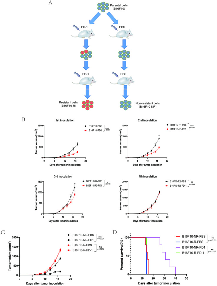
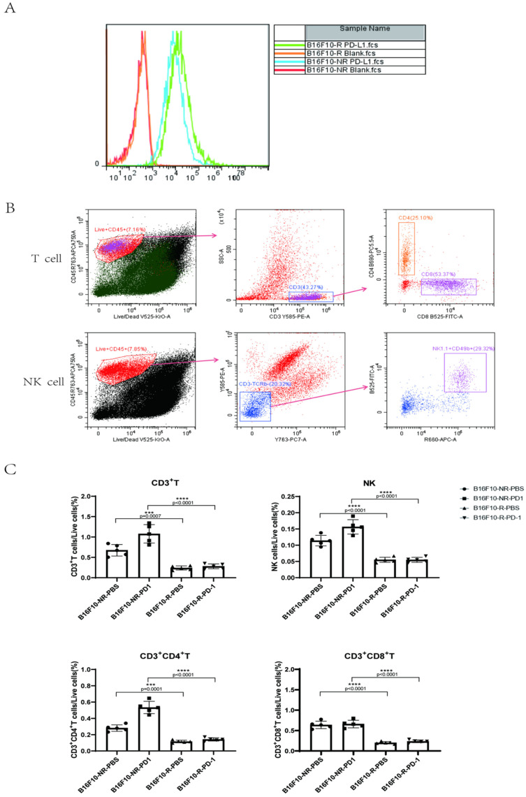
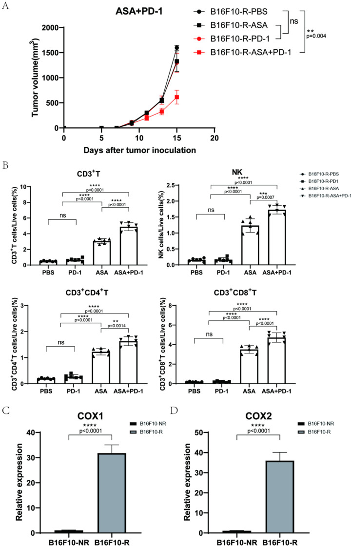
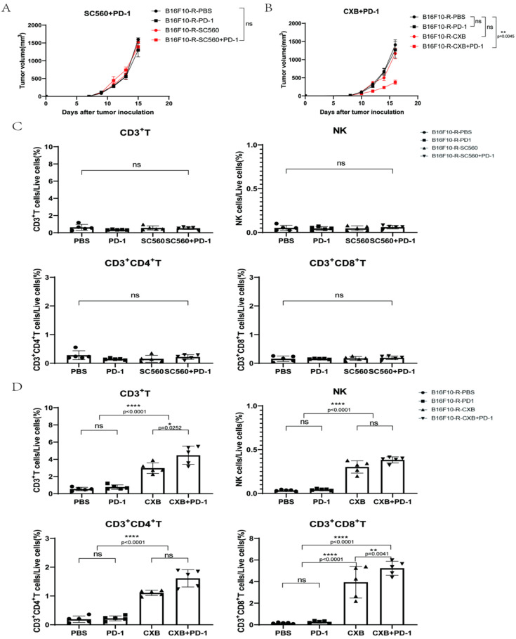
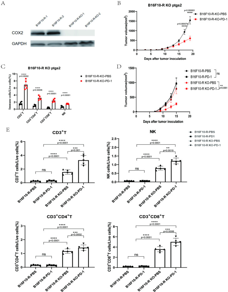
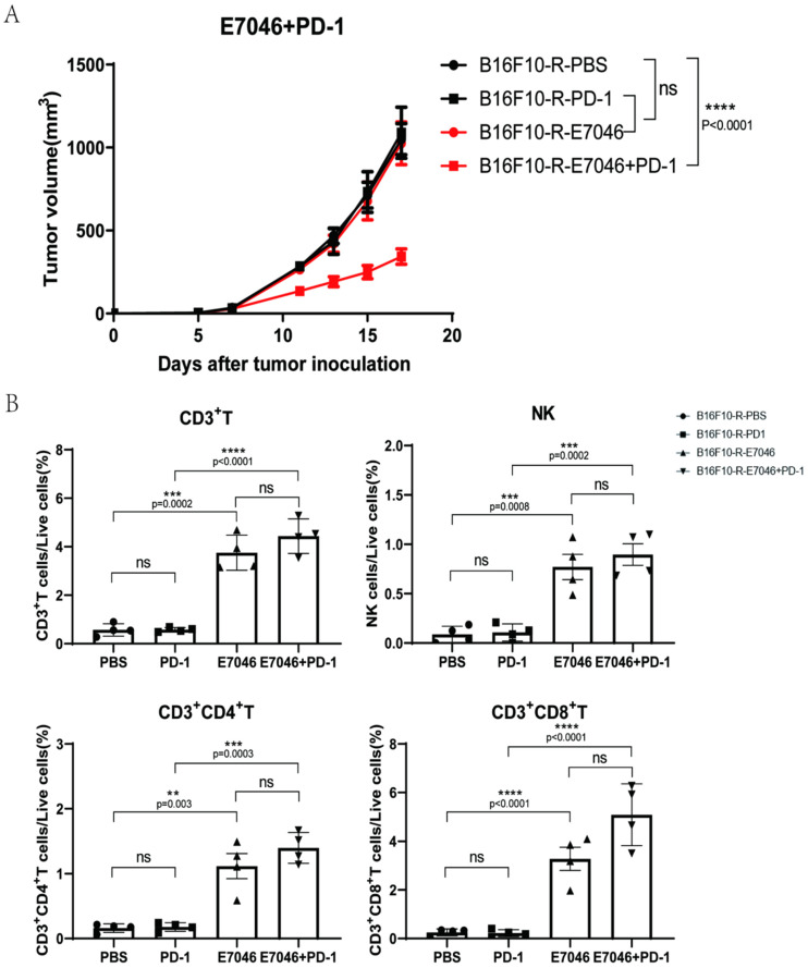
Immunotherapy is an effective method for tumour treatment. Anti-programmed cell death protein 1 (PD-1) and anti-programmed death-ligand 1 (PD-L1) monoclonal antibodies play a significant role in immunotherapy of most tumours; however, some patients develop drug resistance to PD-1/PD-L1 therapy. Cyclooxygenase-2 (COX2) is expressed in various solid tumours, and prostaglandin E2 (PGE2) drives the development of malignant tumours. We developed a drug-resistant B16F10 (B16F10-R) tumour mouse model through four rounds of selection in vivo. Subsequently, we investigated changes in PD-L1 expression and lymphocyte infiltration in B16F10-NR and B16F10-R tumours. Additionally, we explored the role of COX2 in acquired resistance to pembrolizumab, an anti-PD-1 treatment. Immune cell infiltration was significantly decreased in resistant tumours compared to B16F10-NR tumours; however, ptgs2 gene expression was significantly elevated in resistant tumours. Aspirin or celecoxib combined with pembrolizumab can effectively reverse tumour drug resistance. In addition, ptgs2 knockout or the use of the EP4 inhibitor E7046 abrogated drug resistance to anti-PD-1 treatment in B16F10-R tumour cells. Our study showed that inhibition of the COX2/PGE2/EP4 axis could increase the number of immune cells infiltrating the tumour microenvironment and recover drug-resistant tumour sensitivity to pembrolizumab. Thus, we highlight COX2 inhibition as a promising therapeutic target for drug-resistant tumours for future consideration.

Keywords: cyclooxygenase-2; immunosuppression; programmed death-ligand 1; tumour resistance.


Conflict of interest statement

The authors declare no conflict of interest.

Figures

Figure 1
Figure 1
Construction of B16F10 tumour model resistant to anti-PD-1 therapy. (A) Construction method of anti-PD-1-resistant B16F10 tumour cells. (B) Tumour growth curves for four rounds of anti-PD-1-resistant B16F10 tumour selection (n = 6/group, two-way ANOVA test, Sidak). (C) Growth curves of B16F10-NR and B16F10-R tumours. Mice were treated with either pembrolizumab or PBS (n = 6–8/group, two-way ANOVA test, Tukey). (D) Mouse survival curves for B16F10-NR and B16F10-R tumours. Mice were treated with either pembrolizumab or PBS (n = 6–8/group, log-rank test of survival curve); ns, not statistically significant; * p < 0.05; ** p < 0.01; **** p < 0.0001.
Figure 2
Figure 2
Differences between B16F10-NR tumour and B16F10-R tumour cells. (A) PD-L1 expression on the surface of B16F10-NR and B16F10-R cells. (B) Gating strategy to identify intratumoural T and NK cells. (C) Differences in the number of infiltrating lymphocytes in the TME of B16F10-NR tumours and B16F10-R tumours (one-way ANOVA test, Tukey); ns, not statistically significant; *** p < 0.001; **** p < 0.0001.
Figure 3
Figure 3
Effects of aspirin combined with pembrolizumab on B16F10-R tumour growth in vivo. (A) Growth curves of B16F10-R tumours in vivo treated with pembrolizumab, ASA, or PBS (n = 6–8/group, two-way ANOVA test, Tukey). (B) Differences in the number of infiltrating lymphocytes in B16F10-R tumours treated with ASA (one-way ANOVA test, Tukey). (C,D) RT-PCR analysis of ptgs1 (C) and ptgs2 (D) mRNA expression in B16F10-NR and B16F10-R tumours. GAPDH mRNA expression was used as the control (n = 8/group, unpaired, two-tailed t test); ns, not statistically significant; ** p < 0.01; *** p < 0.001; **** p < 0.0001.
Figure 4
Figure 4
Effects of SC560 or celecoxib combined with pembrolizumab on B16F10-R tumour growth in vivo. (A) Growth curves of B16F10-R tumours in vivo treated with pembrolizumab, SC560, or PBS (n = 6–8/group, two-way ANOVA test, Tukey). (B) Growth curves of B16F10-R tumours in vivo treated with pembrolizumab, celecoxib, or PBS (n = 6–8/group, two-way ANOVA test, Tukey). (C) Differences in the number of infiltrating lymphocytes cells in B16F10-R tumours treated with celecoxib (one-way ANOVA test, Tukey). (D) Differences in the number of infiltrating lymphocytes in B16F10-R tumours treated with SC560 (one-way ANOVA test, Tukey); ns, not statistically significant; * p < 0.05; ** p < 0.01; **** p < 0.0001.
Figure 5
Figure 5
COX2 knockout in B16F10-R cells abrogates acquired resistance to anti-PD-1 therapy. (A) Western blot analysis for COX2 expression. GAPDH was the control in the two cell lines. (B) Tumour growth curves of B16F10-R-knockout tumours in vivo treated with pembrolizumab or PBS (n = 6–8/group, two-way ANOVA test, Tukey). (C) Differences in the number of infiltrating lymphocytes in B16F10-R-knockout tumours (one-way ANOVA test, Tukey). (D) Tumour growth curves of B16F10-R and B16F10-R-knockout tumours in vivo treated with pembrolizumab or PBS (n = 6–8/group, two-way ANOVA test, Tukey). (E) Differences in the number of infiltrating lymphocytes in B16F10-R-knockout tumours (one-way ANOVA test, Tukey); ns, not statistically significant; ** p < 0.01; *** p < 0.001; **** p < 0.0001.
Figure 6
Figure 6
Effects of E7046 combined with pembrolizumab on B16F10-R tumour growth in vivo. (A) Growth curves of B16F10-R tumours in vivo treated with pembrolizumab, E7046, or PBS (n = 6–8/group, two-way ANOVA test, Tukey). (B) Differences in the number of infiltrating lymphocytes in B16F10-R tumours treated with E7046 (one-way ANOVA test, Tukey); ns, not statistically significant; ** p < 0.01; *** p < 0.001; **** p < 0.0001.

References

    1. Pardoll D.M. The blockade of immune checkpoints in cancer immunotherapy. Nat. Rev. Cancer. 2012;12:252–264. doi: 10.1038/nrc3239. - DOI - PMC - PubMed
    1. Zou W. Immunosuppressive networks in the tumour environment and their therapeutic relevance. Nat. Rev. Cancer. 2005;5:263–274. doi: 10.1038/nrc1586. - DOI - PubMed
    1. Salmaninejad A., Valilou S.F., Shabgah A.G., Aslani S., Alimardani M., Pasdar A., Sahebkar A. PD-1/PD-L1 pathway: Basic biology and role in cancer immunotherapy. J. Cell. Physiol. 2019;234:16824–16837. doi: 10.1002/jcp.28358. - DOI - PubMed
    1. Frydenlund N., Mahalingam M. PD-L1 and immune escape: Insights from melanoma and other lineage-unrelated malignancies. Hum. Pathol. 2017;66:13–33. doi: 10.1016/j.humpath.2017.06.012. - DOI - PubMed
    1. Zou W., Chen L. Inhibitory B7-family molecules in the tumour microenvironment. Nat. Rev. Immunol. 2008;8:467–477. doi: 10.1038/nri2326. - DOI - PubMed