. 2022 Aug 30;14(17):4208.
doi: 10.3390/cancers14174208.

Demonstrating Tumor Vascular Disrupting Activity of the Small-Molecule Dihydronaphthalene Tubulin-Binding Agent OXi6196 as a Potential Therapeutic for Cancer Treatment

Affiliations

Demonstrating Tumor Vascular Disrupting Activity of the Small-Molecule Dihydronaphthalene Tubulin-Binding Agent OXi6196 as a Potential Therapeutic for Cancer Treatment

Li Liu et al. Cancers (Basel). .

Abstract

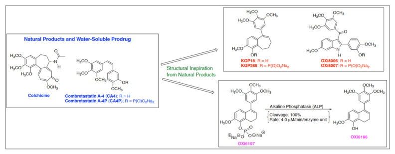
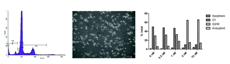
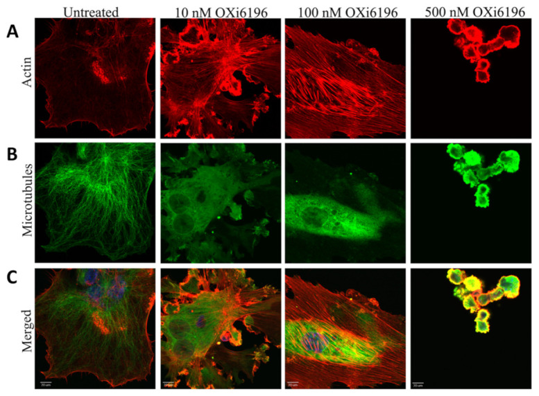
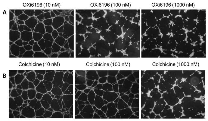
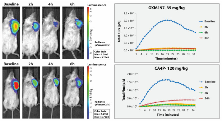
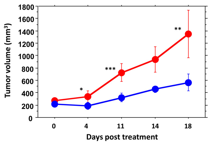
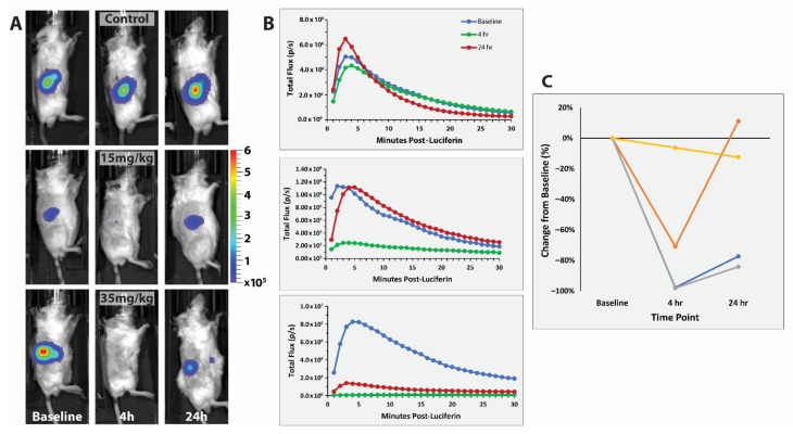
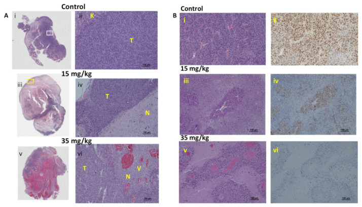
The vascular disrupting activity of a promising tubulin-binding agent (OXi6196) was demonstrated in mice in MDA-MB-231 human breast tumor xenografts growing orthotopically in mammary fat pad and syngeneic RENCA kidney tumors growing orthotopically in the kidney. To enhance water solubility, OXi6196, was derivatized as its corresponding phosphate prodrug salt OXi6197, facilitating effective delivery. OXi6197 is stable in water, but rapidly releases OXi6196 in the presence of alkaline phosphatase. At low nanomolar concentrations OXi6196 caused G2/M cell cycle arrest and apoptosis in MDA-MB-231 breast cancer cells and monolayers of rapidly growing HUVECs underwent concentration-dependent changes in their morphology. Loss of the microtubule structure and increased bundling of filamentous actin into stress fibers followed by cell collapse, rounding and blebbing was observed. OXi6196 (100 nM) disrupted capillary-like endothelial networks pre-established with HUVECs on Matrigel®. When prodrug OXi6197 was administered to mice bearing orthotopic MDA-MB-231-luc tumors, dynamic bioluminescence imaging (BLI) revealed dose-dependent vascular shutdown with >80% signal loss within 2 h at doses ≥30 mg/kg and >90% shutdown after 6 h for doses ≥35 mg/kg, which remained depressed by at least 70% after 24 h. Twice weekly treatment with prodrug OXi6197 (20 mg/kg) caused a significant tumor growth delay, but no overall survival benefit. Similar efficacy was observed for the first time in orthotopic RENCA-luc tumors, which showed massive hemorrhage and necrosis after 24 h. Twice weekly dosing with prodrug OXi6197 (35 mg/kg) caused tumor growth delay in most orthotopic RENCA tumors. Immunohistochemistry revealed extensive necrosis, though with surviving peripheral tissues. These results demonstrate effective vascular disruption at doses comparable to the most effective vascular-disrupting agents (VDAs) suggesting opportunities for further development.

Keywords: bioluminescence imaging (BLI); breast tumors; combretastatin; dihydronaphthalene; kidney tumors; lung tumors; vascular-disrupting agent (VDA).


Conflict of interest statement

The funders had no role in the design of the study; in the collection, analyses or interpretation of the data; in the writing of the manuscript or in the decision to publish the results. K.G.P. holds several patents relevant to the synthesis and use of vascular-disrupting agents.

Figures

Figure 1
Figure 1
Molecular structures of OXi6196 and phosphate prodrug OXi6197, as well as related molecules exhibiting vascular disrupting activity. Exposure of prodrug OXi6197 to alkaline phosphatase leads to rapid release of OXi6196 [27].
Figure 2
Figure 2
Cell cycle arrest. Concentration-dependent G2/M arrest caused by OXi6196 in MDA-MB-231 cells, as assessed by flow cytometry. G2/M blockade was observed at 5 nM with a significant increase in aneuploidy. Cells have started to round and detach. Effects on cell cycle progression and corresponding images are shown in Supplementary material Figures S1 and S2 for OXi6196 and OXi6197, respectively.
Figure 3
Figure 3
Confocal images of endothelial cells fixed and stained for actin and microtubules. Monolayers of rapidly growing HUVECs underwent concentration-dependent changes in the cell morphology with OXi6196 treatment (2 h) that demonstrated an initial loss of the microtubule structure and increased bundling of filamentous actin into stress fibers. Many cells with two nuclei were observed. At higher concentrations (500 nM) cells had detached with remaining adherent cells rounded, condensed and showing multiple blebs. Representative confocal images for endothelial cells fixed and stained with (A) Alexa Fluor™ 594-conjugated phalloidin (red, actin), (B) anti-α-tubulin antibody (green, microtubules), (C) merged image. Bars, 20 μm.
Figure 4
Figure 4
Capillary network disruption. (A) HUVEC capillary-like networks cultured on Matrigel® showed dose-dependent tube disruption with OXi6196 treatment becoming significant at 100 nM. (B) Colchicine ultimately showed similar activity, but at about 10× higher concentration. Cells were imaged with an inverted microscope (5×) and nine fields were photographed per well. A minimum of three independent experiments was carried out for each treatment, and representative images are shown. An example of untreated control is shown in Figure S4.
Figure 5
Figure 5
Comparison of vascular disrupting activity of OXi6197 and CA4P in orthotopic MDA-MB-231-luc tumors. (Right) BLI light emission curves obtained after administering luciferin to mice at various times after VDA. Significantly reduced light emission was observed following administration of VDA. (Left) Heat maps show light emission intensity at about 12 min after administering luciferin overlaid on photographs of mice. Dose response presented in Supplementary Figures S5 and S6.
Figure 6
Figure 6
Growth curves for groups of orthotopic MDA-MB-231-luc tumors comparing saline controls (red, n = 6) and OXi6197 (20 mg/kg twice weekly, blue n = 5). At baseline the two groups had similar mean volumes, but within 4 days of treatment mean tumor volume was significantly different: 4 days (* p < 0.05), day 11 (*** p < 0.001) and day 18 (** p < 0.01). Curves for individual tumors are presented in Supplementary Figure S7.
Figure 7
Figure 7
Vascular disruption assessed in RENCA-luc tumors using dynamic BLI. Effect of OXi6197 on RENCA-luc tumors growing orthotopically in the right kidney of syngeneic BALB/c mice. (A) Top row. Light emission observed from control tumor about 5 min after administering luciferin at baseline and 4 h and 24 h after saline. Middle row) Similar to A, but for a mouse which received OXi6197 (15 mg/kg IP). Bottom row) Similar to B, but for a mouse receiving 35 mg/kg OXi6197. (B) Curves showing evolution of light intensity at various times following OXi6197. Baseline (blue), 4 h (green) and 24 h (red). (C) Comparison of relative signal intensity indicating dose response for four tumors dosed with saline (control), 15 mg/kg and 35 mg/kg (including one small and one large: 0.97 g and 2.7 g, respectively, as determined upon sacrifice and excision).
Figure 8
Figure 8
Histology of RENCA-luc tumors. (A) H&E stained whole mount sections and corresponding magnified regions obtained from RENCA-luc tumors 24 h after administering saline or OXi6197. (i) The control tumor from the mouse receiving saline shows primarily viable tissue with minimal necrosis, (ii) The inset shows tightly packed high-grade tumor cells (T) and remaining normal kidney from site of tumor implantation (K), (iii) tumor exposed to 15 mg/kg OXi6197 showing extensive necrosis (N) and thin viable rim and focal areas of hemorrhage, (iv) expanded view from region in yellow box showing thin rim of viable tumor (T) and extensive necrosis (N), (v) Section from tumor exposed to 35 mg/kg OXi6197 shows massive hemorrhage, and (vi) highly congested blood vessels (v) and extensive necrosis (N). (B) H&E and Ki67 stained sections from the same tumors showing findings associated with vasculature. (i) Control tumor shows densely packed cells and many compressed blood vessels, (ii) The cells show high proliferation activity, (iii) Following 15 mg/kg OXi6197 blood vessels appeared dilated surrounded by rim of viable cells, which (iv) showed retained proliferation, (v) Following 35 mg/kg, vessels appeared dilated and congested, and (vi) surrounding cells appeared less viable, as confirmed by Ki67 staining showing minimal proliferation. Further sections stained for CD31 are shown in Figure S9.
Figure 9
Figure 9
Growth curves for groups of orthotopic RENCA-luc tumors. Saline controls (red, n = 7) are compared with OXi6197 (35 mg/kg twice weekly, blue n = 4; an outlier is excluded from the group presentation). Mean tumor BLI signal as a surrogate for tumor volume was not significantly different at any given time point (p > 0.05), but overall the treated tumors were significantly smaller (based on light emission) than controls over the first 28 days after treatment was initiated (p < 0.003). Compared to baseline, control tumors were significantly larger after 21 days (p = 0.0176), while tumors treated with OXi6197 required 35 days to be significantly larger (p = 0.0035). Curves for individual tumors are presented in Supplementary Figure S10.

References

    1. Siemann D.W. The unique characteristics of tumor vasculature and preclinical evidence for its selective disruption by Tumor-Vascular Disrupting Agents. Cancer Treat. Rev. 2011;37:63–74. doi: 10.1016/j.ctrv.2010.05.001. - DOI - PMC - PubMed
    1. Hinnen P., Eskens F.A. Vascular disrupting agents in clinical development. Br. J. Cancer. 2007;96:1159–1165. doi: 10.1038/sj.bjc.6603694. - DOI - PMC - PubMed
    1. Nepali K., Ojha R., Lee H.Y., Liou J.P. Early investigational tubulin inhibitors as novel cancer therapeutics. Expert Opin. Investig. Drugs. 2016;25:917–936. doi: 10.1080/13543784.2016.1189901. - DOI - PubMed
    1. Siemann D.W., Chaplin D.J., Horsman M.R. Realizing the Potential of Vascular Targeted Therapy: The Rationale for Combining Vascular Disrupting Agents and Anti-Angiogenic Agents to Treat Cancer. Cancer Investig. 2017;35:519–534. doi: 10.1080/07357907.2017.1364745. - DOI - PubMed
    1. Denekamp J. Vascular Attack as a Therapeutic Strategy for Cancer. Cancer Metastasis Rev. 1990;9:267–282. doi: 10.1007/BF00046365. - DOI - PubMed

LinkOut - more resources