Review
. 2022 Sep 2;14(17):4305.
doi: 10.3390/cancers14174305.

Bone Health Management in the Continuum of Prostate Cancer Disease

Affiliations
Review

Bone Health Management in the Continuum of Prostate Cancer Disease

Ettickan Boopathi et al. Cancers (Basel). .

Abstract

Prostate cancer (PCa) is the second-leading cause of cancer-related deaths in men. PCa cells require androgen receptor (AR) signaling for their growth and survival. Androgen deprivation therapy (ADT) is the preferred treatment for patients with locally advanced and metastatic PCa disease. Despite their initial response to androgen blockade, most patients eventually will develop metastatic castration-resistant prostate cancer (mCRPC). Bone metastases are common in men with mCRPC, occurring in 30% of patients within 2 years of castration resistance and in >90% of patients over the course of the disease. Patients with mCRPC-induced bone metastasis develop lesions throughout their skeleton; the 5-year survival rate for these patients is 47%. Bone-metastasis-induced early changes in the bone that proceed the osteoblastic response in the bone matrix are monitored and detected via modern magnetic resonance and PET/CT imaging technologies. Various treatment options, such as targeting osteolytic metastasis with bisphosphonates, prednisone, dexamethasone, denosumab, immunotherapy, external beam radiation therapy, radiopharmaceuticals, surgery, and pain medications are employed to treat prostate-cancer-induced bone metastasis and manage bone health. However, these diagnostics and treatment options are not very accurate nor efficient enough to treat bone metastases and manage bone health. In this review, we present the pathogenesis of PCa-induced bone metastasis, its deleterious impacts on vital organs, the impact of metastatic PCa on bone health, treatment interventions for bone metastasis and management of bone- and skeletal-related events, and possible current and future therapeutic options for bone management in the continuum of prostate cancer disease.

Keywords: androgen receptor; bisphosphonate; castration-resistant prostate cancer; denosumab; osteoblast; osteoclast; prostate cancer; radium-223; taxane.


Conflict of interest statement

The authors declare no conflict of interest.

Figures
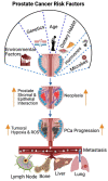
Figure 1
Figure 1
Risk factors in prostate cancer pathogenesis and metastasis. The well-studied prostate cancer risk factors are environmental factors, genetics, age, dietary habits, hormones, and microbial infection. The risk factors increase the ROS levels under tumoral hypoxia and promote stromal epithelial cell interaction and inflammatory signaling molecules that drive tumor initiation. ROS signaling promotes epithelial mesenchymal transition (EMT), and regional and distal metastasis (lymph node, bone, liver, and lung). The orange arrow indicates the increased or progression. This diagrammatic illustration was created with BioRender.com (accessed on 1 August 2022) agreement # OQ23NSL7AN.
Figure 2
Figure 2
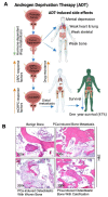
Pathogenesis of prostate cancer bone metastasis and its deleterious impacts in vital organs. (A) The schematic diagram shows that ADT suppresses PCa growth and simultaneously impairs hormone (androgen)-regulated drug metabolizers (cytochrome p450s). Additionally, ADT promotes mental depression, weakness in the bones, heart, lung, and skeletal muscles. ADT also promotes CRPC via AR-dependent and AR-independent mechanisms, drug resistance, and metastatic CRPC (mCRPC). mCRPC promotes local and distal metastasis, poor prognosis, and organ failure. (B) Bright field microscopic image of H&E-stained biopsy samples from (1) a male benign bone PCa (top left), (2) prostate-cancer-induced bone metastasis (top right), (3) prostate-cancer-induced bone metastasis with osteoblastic with woven bone (bottom left), (4) prostate-cancer-induced bone metastasis with osteoblastic bone with calcification (bottom right), magnification 200×. The up-arrow indicates increase and the down arrow indicates decrease. This diagrammatic illustration was created with BioRender.com agreement # CY23NSMIZH.
Figure 3
Figure 3
The impact of metastatic prostate cancer on bone health and bone-metastasis-induced skeletal and non-skeletal events. Prostate cancer often promotes bone metastasis (skeletal metastasis). The prostate cancer cells in the bone interact with bone stromal cells and promote the expression of cytokines, chemokines, growth factors, and osteoclast cell activation. Osteoclast activation initiates bone fracture and calcium release into the blood stream. Excess calcium in the blood stream promotes mental depression, cardiac dysfunction, bone, and muscle weakness, and nephrocalcinosis (kidney disorder) and skeletal and non-skeletal events are managed with lifestyle, nutritional and pharmaceutical interventions. This diagrammatic illustration was created with BioRender.com agreement # JO23NSOQ0.
Figure 4
Figure 4
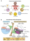
Treatments options for PCa-induced bone metastasis and bone management. (A) Patients with PCa-induced bone metastasis are treated with surgery, hormonal therapy (androgen deprivation therapy, ADT), bone marrow transplant, chemotherapy (taxanes, bisphosphonate), anti-androgen therapy (enzalutamide), personalized medicine (T-cell therapy), radiation therapy (radium-223), and immunotherapy (denosumab). (B) Prostate cancer treatment, including immunotherapy and bone management, is presented in the figure as a schematic diagram. The diagram shows that bisphosphonate reduces osteoclast numbers by promoting apoptosis. Anti-RANKL monoclonal antibody denosumab therapy inhibits the development and activity of osteoclasts, followed by the suppression of bone resorption. This diagrammatic illustration was created with BioRender.com agreement # ET23NSSNDQ and TC23NSPRCC.
Figure 5
Figure 5
Management of PCa bone metastasis induced skeletal-related events (SREs). The cartoon illustrates that changing your lifestyle to a healthy diet with no alcohol and no smoking limits the development of SREs. Weight-bearing exercise for 40 h per week in SRE patients can help to restrict SREs. Boosting immunity also decreases SREs. Pharmacological interventions also help to limit SREs. This diagrammatic illustration was created with BioRender.com agreement # LO23NSQII5.
Figure 6
Figure 6
Multi-omics approach in the identification of potential therapeutic drug targets for prostate cancer. Schematics depict the application of a multi-omics approach in the identification of drug targets. Samples collected from patient-derived (bone mets) xenograft (PDX) (1), PCa-induced lung metastasis (2), and PCa bone mets (3) are subjected to multi-omics analysis. This diagrammatic illustration was created with BioRender.com agreement # JU23NSRKRQ.
Figure 7
Figure 7
Exosomes as a diagnostic tool to identify biomarkers in prostate cancer. Exosomes are non-invasive biomarkers and hold great potential for the diagnosis of prostate cancer. Multi-omics approaches to exosome analysis are summarized in the figure as a schematic diagram. Exosomes are isolated form the blood samples collected from prostate-cancer-induced bone metastatic patients. The non-coding RNA, micro-RNA, proteins, and cell-free DNAs of the exosomes can be identified by RNA, DNA sequencing, 10× spatial genomics, and mass spectrometry. This diagrammatic illustration was created with BioRender.com agreement # IB23NST298.

References

    1. Cuzick J., Thorat M., Andriole G., Brawley O.W., Brown P.H., Culig Z., Eeles R., Ford L.G., Hamdy F.C., Holmberg L., et al. Prevention and early detection of prostate cancer. Lancet Oncol. 2014;15:e484–e492. doi: 10.1016/S1470-2045(14)70211-6. - DOI - PMC - PubMed
    1. Zheng Y., Lin S.X., Wu S., Dahl D.M., Blute M.L., Zhong W.D., Zhou X., Wu C.L. Clinicopathological characteristics of localized prostate cancer in younger men aged ≤ 50 years treated with radical prostatectomy in the PSA era: A systematic review and meta-analysis. Cancer Med. 2020;9:6473–6484. doi: 10.1002/cam4.3320. - DOI - PMC - PubMed
    1. Siegel R.L., Miller K.D., Jemal A. Cancer Statistics. CA Cancer J. Clin. 2017;67:7–30. doi: 10.3322/caac.21387. - DOI - PubMed
    1. Morgenroth A., Cartellieri M., Schmitz M., Günes S., Weigle B., Bachmann M., Abken H., Rieber E.P., Temme A. Targeting of tumor cells expressing the prostate stem cell antigen (PSCA) using genetically engineered T-cells. Prostate. 2007;67:1121–1131. doi: 10.1002/pros.20608. - DOI - PubMed
    1. Rebbeck T.R. Prostate Cancer Genetics: Variation by Race, Ethnicity, and Geography. Semin. Radiat. Oncol. 2016;27:3–10. doi: 10.1016/j.semradonc.2016.08.002. - DOI - PMC - PubMed