. 2022 Aug 31;12(17):2248.
doi: 10.3390/ani12172248.

A Stereological Study of the Three Types of Ganglia of Male, Female, and Undifferentiated Scrobicularia plana (Bivalvia)

Affiliations

A Stereological Study of the Three Types of Ganglia of Male, Female, and Undifferentiated Scrobicularia plana (Bivalvia)

Sukanlaya Tantiwisawaruji et al. Animals (Basel). .

Abstract

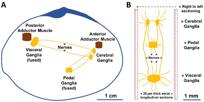
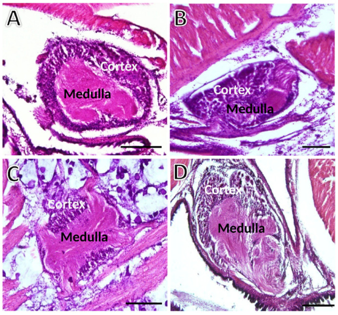
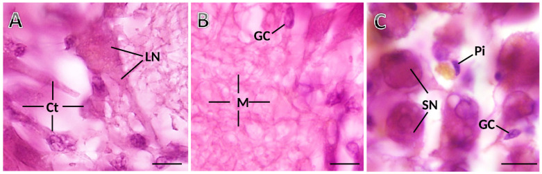
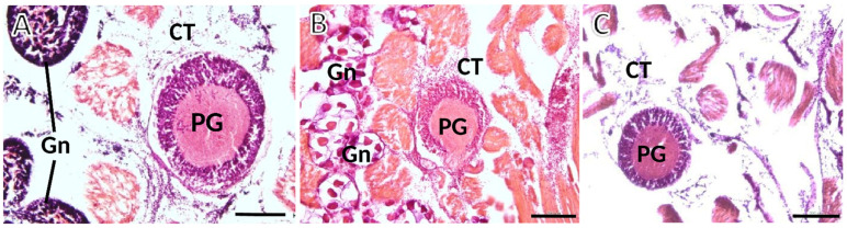
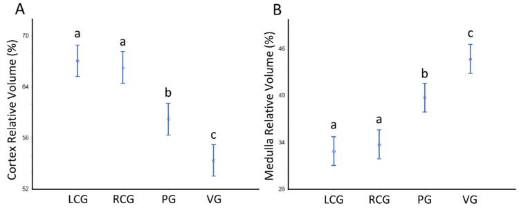
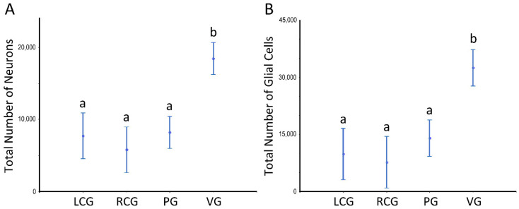
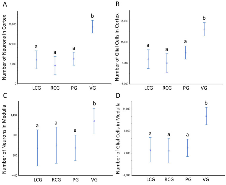
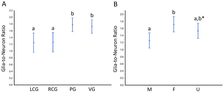
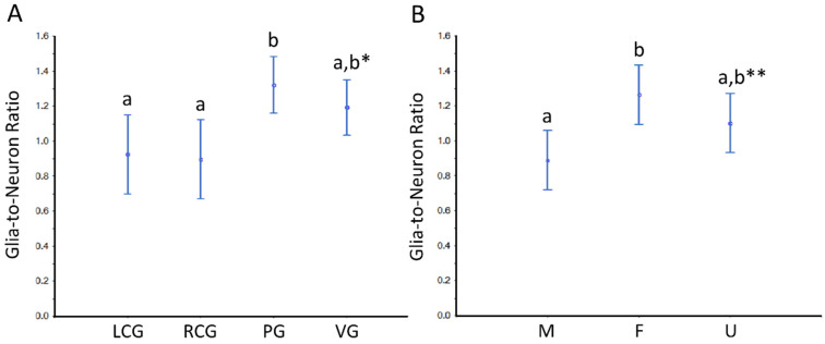
Neurotransmitters modulate gonadal maturation in bivalves. However, it remains unclear whether there are differences in the nervous system structure between sexes, maturation, and ganglia. Therefore, a stereological study was conducted on the ganglia of adult peppery furrow shell (Scrobicularia plana). Equal-sized males, females, and undifferentiated (gamete absence) animals were fixed with 10% formalin and processed for light microscopy. They were serially cut into 35 µm paraffin thick sections and stained with hematoxylin-eosin. Sections with cerebral (cerebropleural), pedal, and visceral ganglia were studied. The parameters estimated were the volumes of the ganglia, the total and relative volumes of their cortex (outer layer) and medulla (neuropil), and the total number of cells (neurons, glia, and pigmented) per ganglia and compartment. The volumes and numbers were estimated, respectively, by the Cavalieri principle and by the optical fractionator. Females show a larger glia to neuron numerical ratio. Further, females have a greater ganglionic volume than undifferentiated adults, with males showing intermediate values. These facts indicate that the ganglia size is related somehow to maturation. The cell size forms the basis of the differences because total cellularity is equal among the groups. The three ganglion types differ in total volumes and the volume ratio of the cortex versus the medulla. The greater volumes of the pedal ganglia (vis-a-vis the cerebral ones) and of the visceral ganglia (in relation to all others) imply more voluminous cortexes and medullae, but more neuronal and non-neuronal cells only in the visceral. The new fundamental data can help interpret bivalve neurophysiology.

Keywords: bivalves; cell numbers; ganglia; glia cells; neurons; sex differences.


Conflict of interest statement

The authors declare no conflict of interest.

Figures

Figure 1
Figure 1
Schematic illustration of the nervous system of S. plana. (A) Longitudinal view of the right valve, depicting the right and left cerebral (cerebropleural) ganglia, pedal ganglia (fused in one), and visceral ganglia (fused in one). (B) Dorsal view of the ganglia, with inter-ganglionic connective nerves and others. The microtome serial sectioning was conducted longitudinally, from right to left.
Figure 2
Figure 2
Transversely sectioned ganglia of S. plana picked randomly for illustration purposes. (A) Left cerebral ganglion. (B) Right cerebral ganglion. (C) Pedal ganglion. (D) Visceral ganglion. The outer basophilic cellular cortex contrasts with the inner medulla. Six animals per sex condition were used. H&E staining. Scale bar = 200 μm.
Figure 3
Figure 3
Photomicrographs taken from thick histological sections of a ganglion of S. plana. (A) Cortex (Ct), with a standing-out large neuron (LN). (B) Medulla (M), with neuronal and glial eosinophilic projections, and somata of glial cells (GC). (C) Detail of cortex, where smaller neurons (SN), one elongated glial cell (GC) and one pigment cell (Pi) are seen. Six animals per sex condition were used. H&E Staining. Scale bar: 10 μm.
Figure 4
Figure 4
Photomicrographs taken from thick histological sections of ganglia of S. plana. (A) Male: spermatogenesis is evident within the gonadal acini (Gn); at the upper and lower left corners of the image. (B) Female: gonadal acini (Gn) are filled with roundish maturing oocytes. (C) Image from one undifferentiated animal, with atrophic acini devoid of maturing gametes, occasionally appearing dispersed in the connective tissue (CT). PG—Pedal ganglion. Six animals per sex were used. H&E staining. Scale bar: 200 μm.
Figure 5
Figure 5
Volumes of the ganglia of S. plana. (A) Per ganglion. (B) All ganglia per sex condition. Different letters mean significant differences (p < 0.05). Data as mean and 95% confidence interval. LCG: left cerebral ganglion; RCG: right cerebral ganglion; PG: pedal ganglion; VG: visceral ganglion; M: males; F: females; U: undifferentiated. Six animals per sex condition were used.
Figure 6
Figure 6
Volumes of the ganglia cortex and medulla of S. plana. (A) Cortex per ganglion. (B) All cortexes per sex condition. (C) Medulla per ganglion. (D) All medullae per sex condition. Different letters mean significant differences (p < 0.05). Data as mean and 95% confidence interval. LCG, RCG, PG, VG, M, F, and U as in Figure 5. * Trend toward a significant difference between M and F (p ≈ 0.05). Six animals per sex condition were used.
Figure 7
Figure 7
Relative volumes (%) of the cerebral, pedal and visceral ganglia cortex and medulla of S. plana, with all sex conditions combined. (A) VV (cortex, ganglion). (B) VV (medulla, ganglion). Different letters mean significant differences (p < 0.05). Data as mean and 95% confidence interval. LCG: left cerebral ganglion; RCG: right cerebral ganglion; PG: pedal ganglia; VG: visceral ganglia. Six animals per sex condition were used.
Figure 8
Figure 8
Number of neurons and glial cells in the ganglia of S. plana; data from all sex conditions combined. (A) Neurons. (B) Glial cells. Different letters mean significant differences. Data as mean and 95% confidence interval. LCG: left cerebral ganglion; RCG: right cerebral ganglion; PG: pedal ganglion; VG: visceral ganglion. Six animals per sex condition were used.
Figure 9
Figure 9
Number of neurons and glial cells in the cortex and medulla of ganglia of S. plana; data from all sex conditions combined. (A) Neurons in the cortex. (B) Glial cells in the cortex. (C) Neurons in the medulla. (D) Glial cells in the medulla. Different letters mean significant differences. Data as mean and 95% confidence interval (p < 0.05). LCG: left cerebral ganglion; RCG: right cerebral ganglion; PG: pedal ganglion; VG: visceral ganglion. Six animals per sex condition were used.
Figure 10
Figure 10
Glia-to-neuron (number) ratio in the ganglia of S. plana, considering the whole ganglia. (A) Data per ganglion type, irrespective of sex condition. (B) Results from all ganglia, grouped per sex condition. Different letters mean significant differences (p < 0.05). Data as mean and 95% confidence interval. LCG: left cerebral ganglion; RCG: right cerebral ganglion; PG: pedal ganglion; VG: visceral ganglion; M: males; F: females; U: undifferentiated. * Trend towards a significant difference between M and U (p ≈ 0.05). Six animals per sex condition were used.
Figure 11
Figure 11
Glia-to-neuron (number) ratio in the cortex of the ganglia of S. plana. (A) Data per ganglion type, irrespective of sex condition. (B) Results from all ganglia, grouped per sex condition. Different letters mean significant differences (p < 0.05). Data as mean and 95% confidence interval. LCG: left cerebral ganglion; RCG: right cerebral ganglion; PG: pedal ganglion; VG: visceral ganglion; M: males; F: females; U: undifferentiated. * Trend toward a significant difference between VG and both LCG and RGC (p ≈ 0.05). ** Trend towards a significant difference between M and U (p ≈ 0.05). Six animals per sex condition were used.

Similar articles

References

    1. Cosgrove K.P., Mazure C.M., Staley J.K. Evolving knowledge of sex differences in brain structure, function and chemistry. Biol. Psychiatry. 2007;62:847–855. doi: 10.1016/j.biopsych.2007.03.001. - DOI - PMC - PubMed
    1. Fowler C., Goerzen D., Madularum D., Devenyim G.A., Chakravarty M.M., Near J. Longitudinal characterization of neuroanatomical changes in the Fischer 344 rat brain during normal aging and between sexes. Neurobiol. Aging. 2022;109:216–228. doi: 10.1016/j.neurobiolaging.2021.10.003. - DOI - PubMed
    1. Zhai G., Jia J., Bereketoglu C., Yin Z., Pradhan A. Sex-specific differences in zebrafish brains. Biol. Sex Differ. 2022;13:31. doi: 10.1186/s13293-022-00442-2. - DOI - PMC - PubMed
    1. Arnold A.P. The gender of the voice within: The neural origin of sex differences in the brain. Curr. Res. Neurobiol. 2003;13:759–764. doi: 10.1016/j.conb.2003.10.005. - DOI - PubMed
    1. Qureshi I.A., Mehler M.F. Genetic and epigenetic underpinnings of sex differences in the brain and in neurological and psychiatric disease susceptibility. Prog. Brain Res. 2010;186:77–95. - PMC - PubMed

LinkOut - more resources