. 2022 Sep 2;12(17):2272.
doi: 10.3390/ani12172272.

Effects of Para-Toluenesulfonamide on Canine Melanoma Xenotransplants in a BALB/c Nude Mouse Model

Affiliations

Effects of Para-Toluenesulfonamide on Canine Melanoma Xenotransplants in a BALB/c Nude Mouse Model

Chien-Teng Lin et al. Animals (Basel). .

Abstract

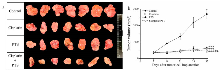
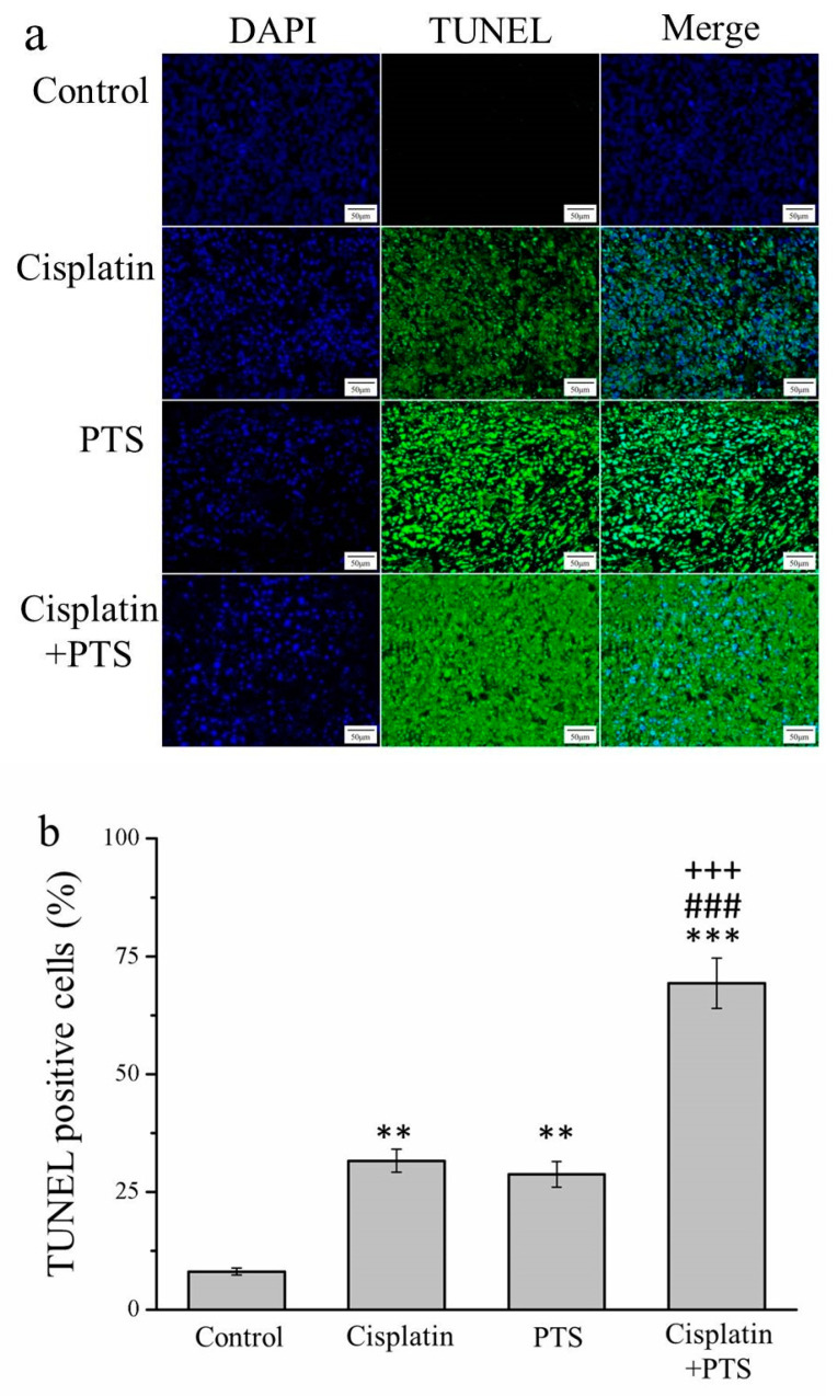
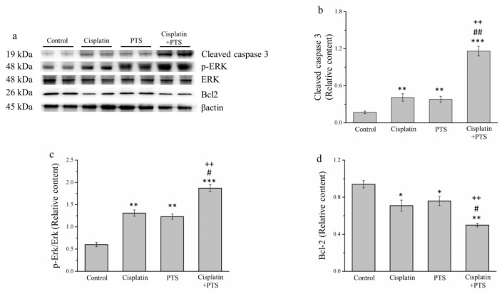
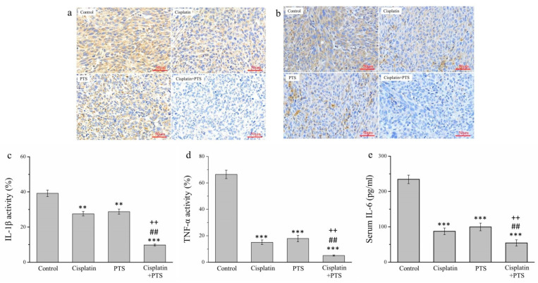
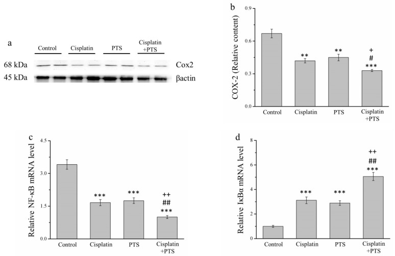
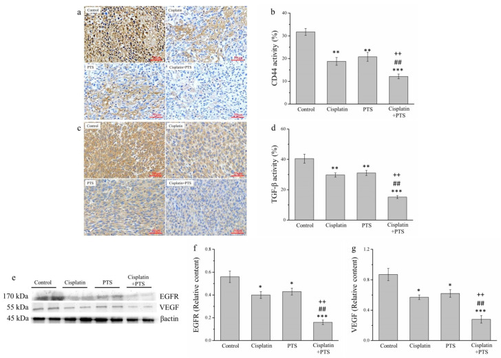
The pharmacological pathway of para-toluenesulfonamide (PTS) restricts the kinase activity of the mammalian target of rapamycin, potentially leading to reductions in cell division, cell growth, cell proliferation, and inflammation. These pathways have a critical effect on tumorigenesis. We aimed to examine the antitumor effect of PTS or PTS combined with cisplatin on canine melanoma implanted in BALB/c nude mice by estimating tumor growth, apoptosis expression, inflammation, and metastasis. The mice were randomly divided into four groups: control, cisplatin, PTS, and PTS combined with cisplatin. Mice treated with PTS or PTS combined with cisplatin had retarded tumor growth and increased tumor apoptosis through the enhanced expression of cleaved caspase 3 and extracellular signal-regulated kinase phosphorylation, decreased inflammatory cytokine levels, reduced inflammation-related factors, enhanced anti-inflammation-related factors, and inhibition of metastasis-related factors. Mice treated with PTS combined with cisplatin exhibited significantly retarded tumor growth, reduced tumor size, and increased tumor inhibition compared with those treated with cisplatin or PTS alone. PTS or PTS combined with cisplatin could retard canine melanoma growth and inhibit tumorigenesis. PTS and cisplatin were found to have an obvious synergistic tumor-inhibiting effect on canine melanoma. PTS alone and PTS combined with cisplatin may be antitumor agents for canine melanoma treatment.

Keywords: apoptosis; canine melanoma; cisplatin; inflammation; metastasis; para-toluenesulfonamide.


Conflict of interest statement

The authors declare no conflict of interest.

Figures

Figure 1
Figure 1
Effects of treatment with cisplatin, para-toluenesulfonamide (PTS), and their combination on M5 canine melanoma tumor growth in BALB/c nude mice. (a) Tumors excised from the mice 35 days after tumor-cell implantation. (b) Change in tumor volume over time. Treatments were administered from day 7 (when the tumors were detected) three times per week, as follows: saline (control), 2 mg/kg cisplatin (cisplatin), 100 mg/kg PTS (PTS), and 100 mg/kg PTS combined with 2 mg/kg cisplatin (cisplatin + PTS). Data are presented as mean ± standard deviation (SD), n = 7 per group. *** p < 0.001 vs. control; # p < 0.05 vs. cisplatin; + p < 0.05 vs. PTS.
Figure 2
Figure 2
TUNEL/DAPI assay of apoptosis in M5 canine melanoma tumors implanted in BALB/c nude mice. (a) Staining with DAPI, TUNEL, and their merging. (b) Percentage of cells in the tumors that were TUNEL-positive in control mice and those administered with one of the following treatments three times per week, starting on day 7 (when the tumors were detected): saline (control), 2 mg/kg cisplatin (cisplatin), 100 mg/kg PTS (PTS), or 100 mg/kg PTS and 2 mg/kg cisplatin (cisplatin + PTS). Data are presented as mean ± SD, n = 7 per group. ** p < 0.01, *** p < 0.001 vs. control; ### p < 0.001 vs. cisplatin; +++ p < 0.001 vs. PTS. Scale bars: 50 µm; magnification: 200×.
Figure 3
Figure 3
Western blot analysis of apoptosis-related proteins in M5 canine melanoma tumors implanted in BALB/c nude mice. (a) Representative Western blot. (bd) Quantitative comparisons of the expression of cleaved caspase 3 (b), phosphorylated ERK (c), and (d) Bcl-2. The mice were administered the following treatments three times per week: saline (control), 2 mg/kg cisplatin (cisplatin), 100 mg/kg PTS (PTS), or 100 mg/kg PTS and 2 mg/kg cisplatin (cisplatin + PTS). Data are presented as mean ± SD, n = 7 per group. * p < 0.05, ** p < 0.01, and *** p < 0.001 vs. control; # p < 0.05 and ## p < 0.01 vs. cisplatin; ++ p < 0.01 vs. PTS.
Figure 4
Figure 4
Immunohistochemical (IHC) analysis of the expression of three inflammatory cytokines in M5 canine melanoma tumors implanted in BALB/c nude mice. (a,b) Representative images showing IL-1β (a) and TNF-α (b) expression. (ce) Quantitative analyses of the IHC expression of IL-1β (c), TNF-α (d), and serum IL-6 (e). The mice were administered the following treatments three times per week: saline (control), 2 mg/kg cisplatin (cisplatin), 100 mg/kg PTS (PTS), or 100 mg/kg PTS and 2 mg/kg cisplatin (cisplatin + PTS). Data are presented as mean ± SD, n = 7 per group. ** p < 0.01 and *** p < 0.001 vs. control; ## p < 0.01 vs. cisplatin; ++ p < 0.01 vs. PTS. Scale bars: 50 µm.
Figure 5
Figure 5
Analysis of factors related to inflammation in M5 canine melanoma tumors implanted in BALB/c nude mice. (a) Representative Western blot of cyclooxygenase-2 (COX-2). (b) Quantitative comparison of COX-2 expression in the four treatment groups. We conducted a quantitative polymerase chain reaction (PCR) to assess the relative mRNA expression levels of (c) NF-κB and (d) IκBα. The mice were administered the following treatments three times per week: saline (control), 2 mg/kg cisplatin (cisplatin), 100 mg/kg PTS (PTS), or 100 mg/kg PTS and 2 mg/kg cisplatin (cisplatin + PTS). Data are presented as mean ± SD, n = 7 per group. ** p < 0.01 and *** p < 0.001 vs. control; # p < 0.05 and ## p < 0.01 vs. cisplatin; + p < 0.05 and ++ p < 0.01 vs. PTS.
Figure 6
Figure 6
Analysis of factors related to metastasis in M5 canine melanoma tumors implanted in BALB/c nude mice. (ad) Representative images (a,c) and quantitative comparisons (b,d) from our IHC analysis of CD44 (a,b) and TGF-β (c,d) expression. (eg) Representative Western blot (e) from our analysis of the relative expression of EGFR (f) and VEGF (g). The mice were administered the following treatments three times per week: saline (control), 2 mg/kg cisplatin (cisplatin), 100 mg/kg PTS (PTS), or 100 mg/kg PTS and 2 mg/kg cisplatin (cisplatin + PTS). Data are presented as mean ± SD, n = 7 per group. * p < 0.05, ** p < 0.01, and *** p < 0.001 vs. control; ## p < 0.01 vs. cisplatin; ++ p < 0.01 vs. PTS. Scale bars: 50 µm.

Similar articles

Cited by

References

    1. Boria P.A., Murry D.J., Bennett P.F., Glickman N.W., Snyder P.W., Merkel B.L., Schlittler D.L., Mutsaers A.J., Thomas R.M., Knapp D.W. Evaluation of cisplatin combined with piroxicam for the treatment of oral malignant melanoma and oral squamous cell carcinoma in dogs. J. Am. Vet. Med. Assoc. 2004;224:388–394. doi: 10.2460/javma.2004.224.388. - DOI - PubMed
    1. Bergman P.J., Kent M.S., Farese J.P. Melanoma. In: Withrow S., Vail D., Page R., editors. Withrow and MacEwen’s Small Animal Clinical Oncology. Elsevier Saunders; St. Louis, MO, USA: 2013. pp. 321–333.
    1. Almela R.M., Ansón A. A Review of Immunotherapeutic Strategies in Canine Malignant Melanoma. Vet. Sci. 2019;6:15–27. - PMC - PubMed
    1. Veena P., Kokila S., Rayadurgam V., Kumar S., Sankar P., Dhanalakshmi N. Malignant melanoma in a Dog—A Case report. Vet. World. 2012;5:431–432. doi: 10.5455/vetworld.2012.431-432. - DOI
    1. Atherton M.J., Morris J.S., McDermott M.R., Lichty B.D. Cancer immunology and canine malignant melanoma: A comparative review. Vet. Immunol. Immunopathol. 2016;169:15–26. - PubMed