

A Novel Muco-Gingival Approach for Immediate Implant Placement to Obtain Soft- and Hard-Tissue Augmentation
- PMID: 36078914
- PMCID: PMC9456498
- DOI: 10.3390/jcm11174985
A Novel Muco-Gingival Approach for Immediate Implant Placement to Obtain Soft- and Hard-Tissue Augmentation
Abstract
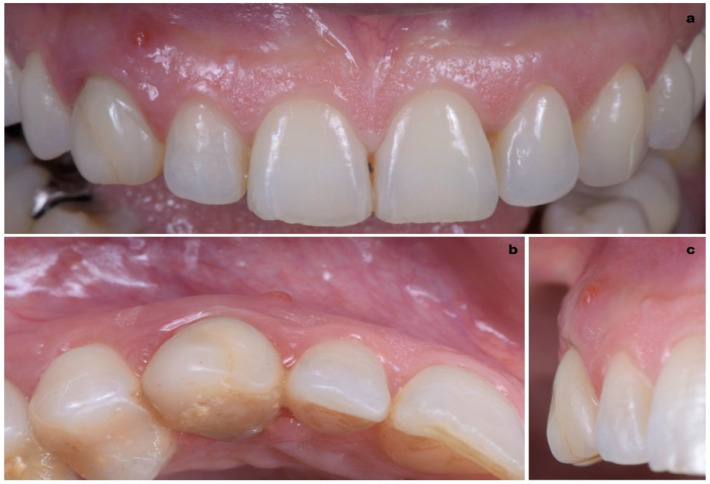
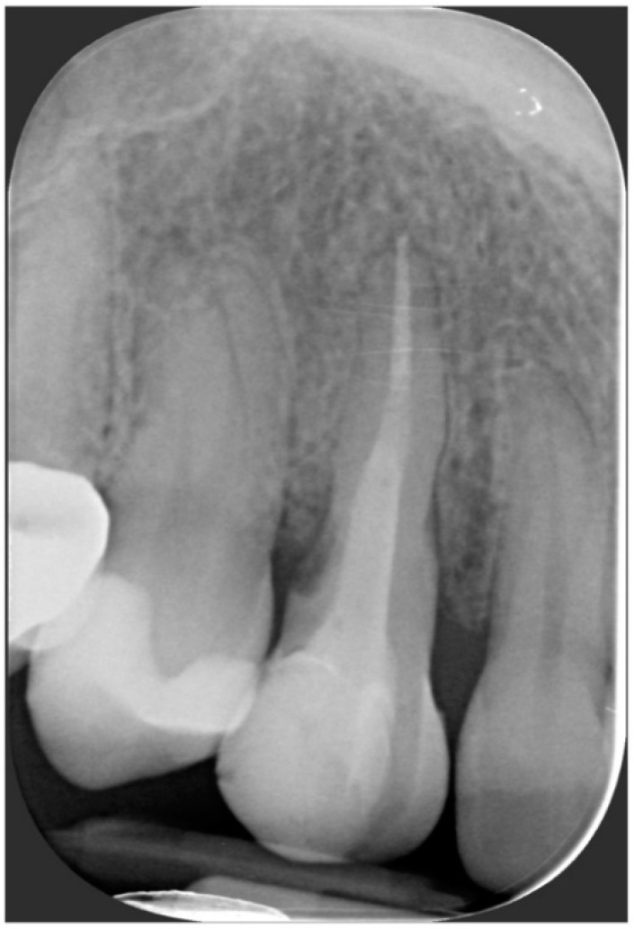
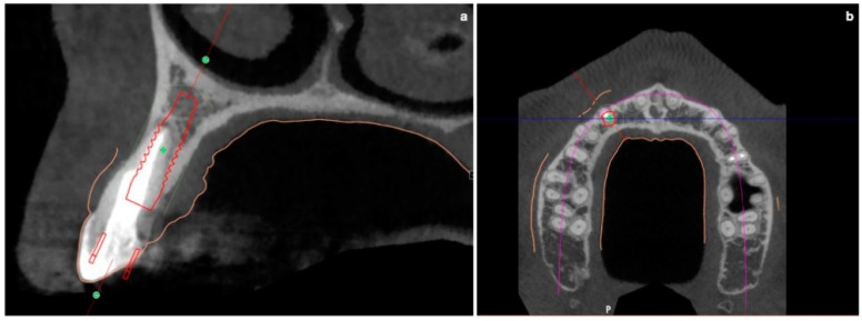
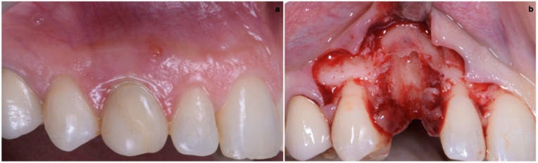
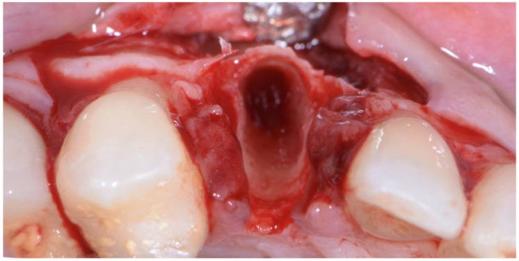
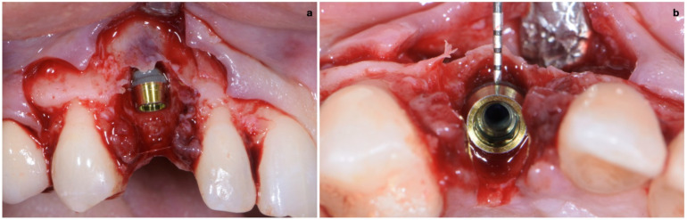
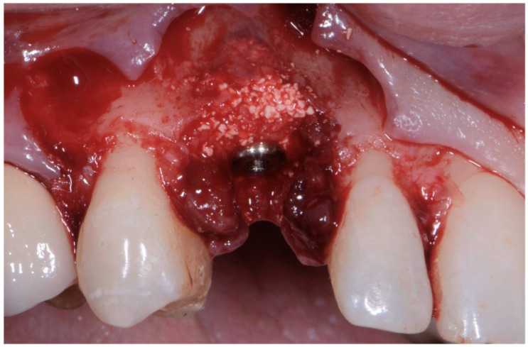
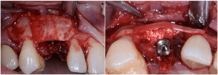
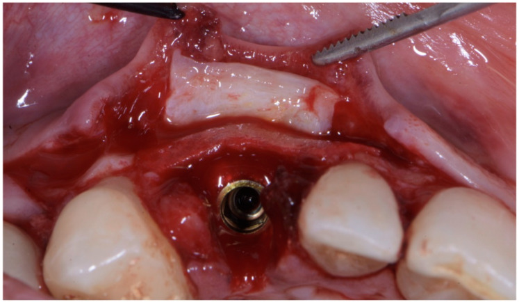
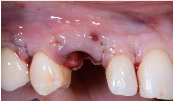
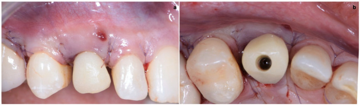
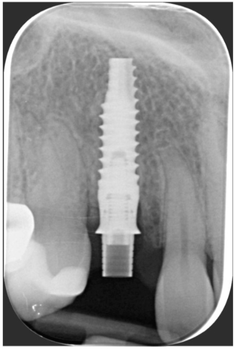
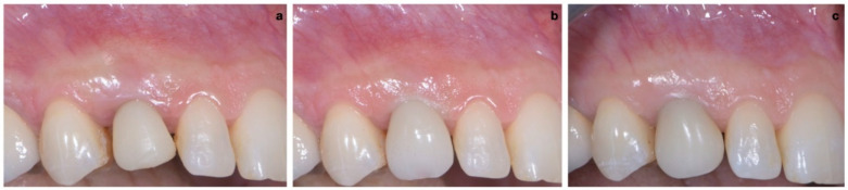
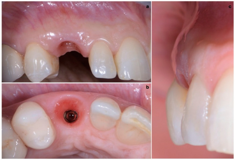
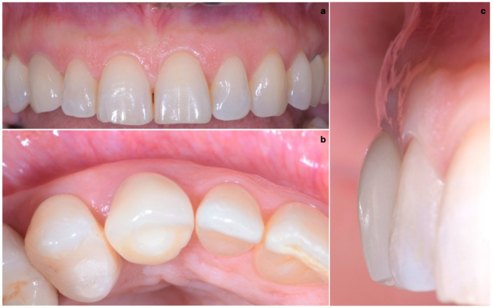
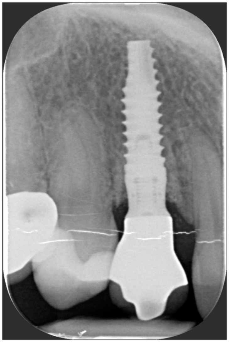
The aim of this article is to describe a novel approach combining muco-gingival, regenerative and prosthetics concepts for immediate implant insertion that overcomes the limits traditionally considered as contraindications for Type 1 flapless implant positioning, simultaneously obtaining soft- and hard-tissue augmentation. After pre-surgical CBCT evaluation, the surgical technique consisted in the execution of a lateral-approach coronally advanced envelope flap, with oblique submarginal interproximal incisions directed towards the flap's center of rotation (the tooth to be extracted); after buccal-flap elevation, the atraumatic extraction of the tooth was performed. Following guided implant insertion, a mixture of biomaterial and autologous bone was placed, stabilized by a pericardium membrane and a connective-tissue graft sutured in the inner aspect of the buccal flap. The peri-implant soft tissues were conditioned with a provisional crown until the shape and position for the mucosal scallop to resemble the gingival margin of the adjacent corresponding tooth were obtained; then, the definitive screw-retained restoration was placed. Within the limitations of this case report, the proposed immediate implant placement approach combining CTG application and buccal bone regeneration showed the possibility of obtaining 1-year-follow-up implant success, stable bone level, good esthetic results and high patient satisfaction.
Keywords: connective-tissue graft; hard-tissue augmentation; immediate implant placement; muco-gingival approach; soft-tissue augmentation.
Conflict of interest statement
The authors declare no conflict of interest.
Figures
References
-
- Nevins M., Wang H. Soft tissue management to augment implant success. In: Zucchelli G., Stefanini M., editors. Implant Therapy: Clinical Approaches and Evidence of Success. Quintessence Publishing; Batavia, IL, USA: 2019. pp. 367–394.
-
- Hartlev J., Kohberg P., Ahlmann S., Andersen N.T., Schou S., Isidor F. Patient satisfaction and esthetic outcome after immediate placement and provisionalization of single-tooth implants involving a definitive individual abutment. Clin. Oral Implants Res. 2014;25:1245–1250. doi: 10.1111/clr.12260. - DOI - PubMed
-
- Khzam N., Arora H., Kim P., Fisher A., Mattheos N., Ivanovski S. Systematic review of soft tissue alterations and esthetic outcomes following immediate implant placement and restoration of single implants in the anterior maxilla. J. Periodontol. 2015;86:1321–1330. doi: 10.1902/jop.2015.150287. - DOI - PubMed
Publication types
LinkOut - more resources
Full Text Sources
