. 2022 Aug 27;11(17):5041.
doi: 10.3390/jcm11175041.

Influence of Radiotherapy on Ossification of Vascularized Osseous Reconstruction of the Jaw: A Radiological Retrospective Cohort Study Based on Panoramic Radiographs

Affiliations

Influence of Radiotherapy on Ossification of Vascularized Osseous Reconstruction of the Jaw: A Radiological Retrospective Cohort Study Based on Panoramic Radiographs

Maximilian Gottsauner et al. J Clin Med. .

Abstract

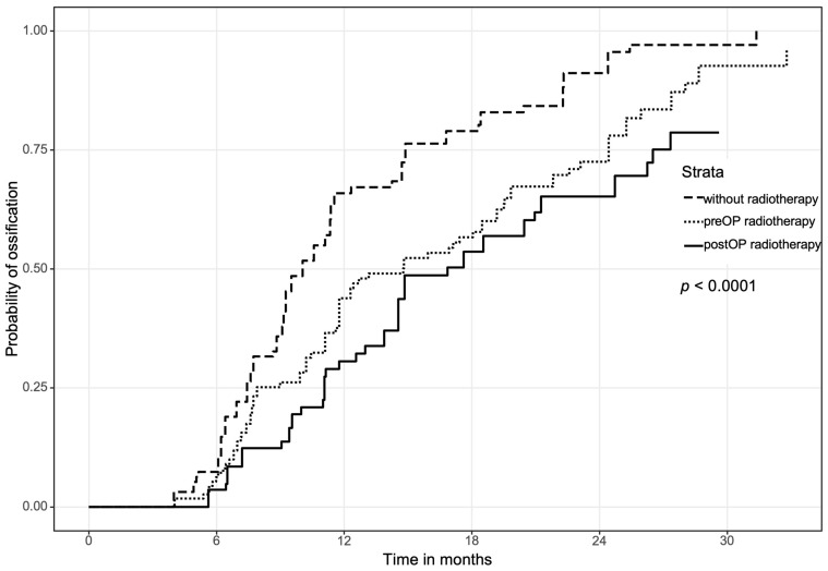
Background: The aim of this study was to evaluate the impact of irradiation and time of irradiation on the ossification of jaws reconstructed with free bone grafts. Methods: In total, 100 reconstructions of the jaw were retrospectively evaluated for ossification between bone segments by two raters based on postoperative panoramic radiographs (immediate postOP, approximately 6, 12 and 24 months follow-up). Three subgroups were divided according to the time of irradiation: preoperative radiation therapy (n = 41), postoperative radiation therapy (n = 26) and patients without any radiation therapy (n = 33) as the control group. Ossification time and influencing factors were documented. Results: The fastest ossification with a median of 304 ± 37 days was observed (p < 0.001) in the nonirradiated control group. No significant difference (p = 0.087) in ossification was found between the pre- (447 ± 136 days) and postoperative (510 ± 112 days) radiation groups. Ossification between two graft segments (336 ± 38 days) showed significantly (p < 0.001) faster ossification than between the original and grafted bone (448 ± 85 days). Moreover, closer initial contact between the segments resulted in faster ossification (p < 0.001). When analyzing cofactors, tobacco consumption was the only negative factor aggravating ossification (p = 0.006). Conclusion: Head and neck radiation corresponded with the impaired and prolonged ossification of jaw reconstructions with free bone grafts. There was no difference in ossification if radiotherapy was performed before or after reconstructive surgery. A close bony contact was particularly important for ossification between the original and grafted bone.

Keywords: fibula; jaw; mandible; microvascular reconstruction; ossification; radiotherapy.


Conflict of interest statement

The authors declare no conflict of interest.

Figures

Figure 1
Figure 1
Various examples for ossification in different radiographs: (a,b) show patient after preoperative radiotherapy, consecutive osteoradionecrosis of the mandible and reconstruction with a 3-segmented fibula. (a) Initial postsurgical radiograph with good contact between fibular segments in left canine area, moderate contact in right canine and right angle area and no contact in left angle area. (b) After 6 months, complete intersegmental ossification and nonossification between segments and original bone. (c,d) show young patient after gunshot wound and reconstruction with a two-segmental fibula. (c) Initial postsurgical radiograph with good contact between fibula segments and graft/native bone. (d) After 1 year, complete ossification of all contacts allowed for removal of the reconstruction plate.
Figure 2
Figure 2
Overall complete ossification for every point of contact depending on radiotherapy.
Figure 3
Figure 3
(a) Ossification concerning the contact between two segments of the transplant and between one segment of the transplant and the original bone of the jaw. (b) Ossification concerning the contact between two segments of the transplant and between one segment of the transplant and the original bone, subdivided regarding time and event of radiation therapy. Bone healing was faster between graft segments compared to segment/jaw contacts in both preORT and postORT groups.
Figure 3
Figure 3
(a) Ossification concerning the contact between two segments of the transplant and between one segment of the transplant and the original bone of the jaw. (b) Ossification concerning the contact between two segments of the transplant and between one segment of the transplant and the original bone, subdivided regarding time and event of radiation therapy. Bone healing was faster between graft segments compared to segment/jaw contacts in both preORT and postORT groups.
Figure 4
Figure 4
(a) Overall locations of the mandible. Reconstructions of the maxilla were excluded in this figure due to a lack of data. (b) Separate evaluation of the contralateral (opposite) side. Significantly worse ossification in the postORT group.
Figure 4
Figure 4
(a) Overall locations of the mandible. Reconstructions of the maxilla were excluded in this figure due to a lack of data. (b) Separate evaluation of the contralateral (opposite) side. Significantly worse ossification in the postORT group.

References

    1. Cannady S.B., Lamarre E., Wax M.K. Microvascular Reconstruction: Evidence-Based Procedures. Facial Plast. Surg. Clin. N. Am. 2015;23:347–356. doi: 10.1016/j.fsc.2015.04.007. - DOI - PubMed
    1. Largo R.D., Garvey P.B. Updates in Head and Neck Reconstruction. Plast. Reconstr. Surg. 2018;141:271e–285e. doi: 10.1097/PRS.0000000000004070. - DOI - PubMed
    1. Al Deek N.F. Osteoradionecrosis of the mandible: Why not to be more aggressive in earlier stage? Am. J. Otolaryngol. 2020;41:102343. doi: 10.1016/j.amjoto.2019.102343. - DOI - PubMed
    1. Haroun K., Coblens O.M. Reconstruction of the mandible for osteoradionecrosis. Curr. Opin. Otolaryngol. Head Neck Surg. 2019;27:401–406. doi: 10.1097/MOO.0000000000000571. - DOI - PubMed
    1. Goh B.T., Lee S., Tideman H., Stoelinga P.J. Mandibular reconstruction in adults: A review. Int. J. Oral Maxillofac. Surg. 2008;37:597–605. doi: 10.1016/j.ijom.2008.03.002. - DOI - PubMed

LinkOut - more resources