. 2022 Aug 28;11(17):5048.
doi: 10.3390/jcm11175048.

The Effects of Scanning Speed and Standoff Distance of the Fiber on Dusting Efficiency during Short Pulse Holmium: YAG Laser Lithotripsy

Affiliations

The Effects of Scanning Speed and Standoff Distance of the Fiber on Dusting Efficiency during Short Pulse Holmium: YAG Laser Lithotripsy

Junqin Chen et al. J Clin Med. .

Abstract

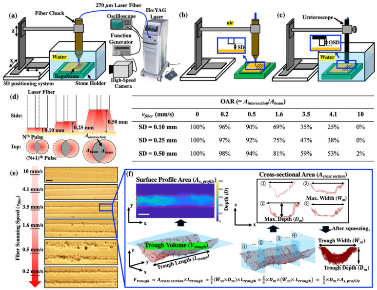
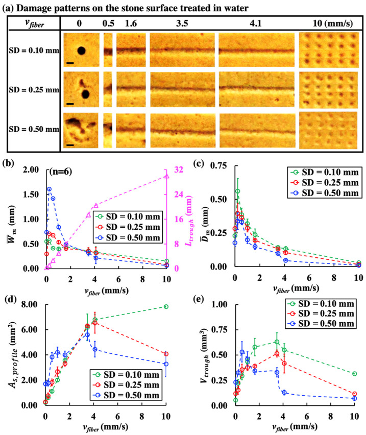
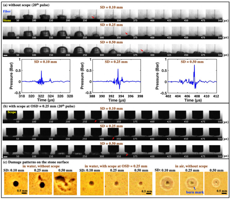
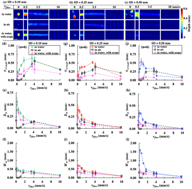
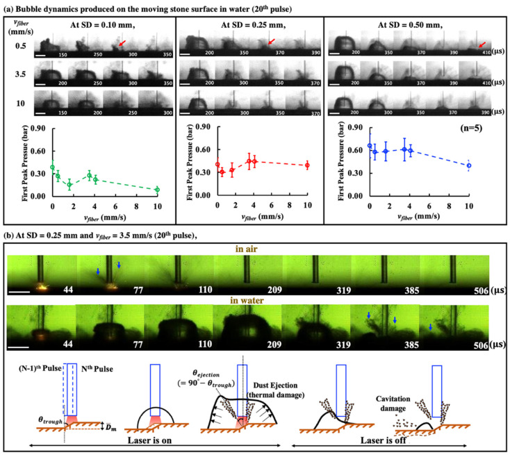
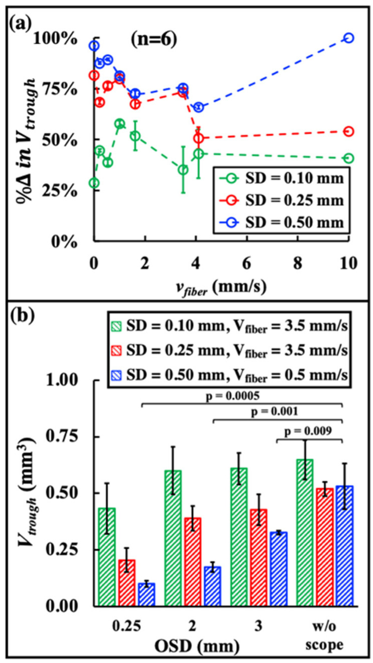
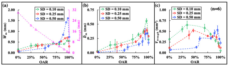
To investigate the effects of fiber lateral scanning speed across the stone surface (vfiber) and fiber standoff distance (SD) on dusting efficiency during short pulse holmium (Ho): YAG laser lithotripsy (LL), pre-soaked BegoStone samples were treated in water using 0.2 J/20 Hz at SD of 0.10~0.50 mm with vfiber in the range of 0~10 mm/s. Bubble dynamics, pressure transients, and stone damage were analyzed. To differentiate photothermal ablation vs. cavitation damage, experiments were repeated in air, or in water with the fiber tip at 0.25 mm proximity from the ureteroscope end to mitigate cavitation damage. At SD = 0.10 mm, the maximum dusting efficiency was produced at vfiber = 3.5 mm/s, resulting in long (17.5 mm), shallow (0.15 mm), and narrow (0.4 mm) troughs. In contrast, at SD = 0.50 mm, the maximum efficiency was produced at vfiber = 0.5 mm/s, with much shorter (2.5 mm), yet deeper (0.35 mm) and wider (1.4 mm), troughs. With the ureteroscope end near the fiber tip, stone damage was significantly reduced in water compared to those produced without the ureteroscope. Under clinically relevant vfiber (1~3 mm/s), dusting at SD = 0.5 mm that promotes cavitation damage may leverage the higher frequency of the laser (e.g., 40 to 120 Hz) and, thus, significantly reduces the procedure time, compared to at SD = 0.1 mm that promotes photothermal ablation. Dusting efficiency during short pulse Ho: YAG LL may be substantially improved by utilizing an optimal combination of vfiber, SD, and frequency.

Keywords: cavitation; fiber scanning speed; laser lithotripsy; mechanisms of stone dusting.


Conflict of interest statement

The authors declare no conflict of interest. The funders had no role in the design of the study; in the collection, analyses, or interpretation of data; in the writing of the manuscript; or in the decision to publish the results.

Figures

Figure 1
Figure 1
Experimental setup for pre-soaked BegoStone samples treated with perpendicular fiber (core diameter = 270 mm) placed at different fiber tip-to-stone standoff distances (SDs) (a) in water, synced with high-speed imaging from the side-view, (b) in air, and (c) in water with a ureteroscope placed at various offset distances (OSDs). A closer view of the gaps between the scope end, fiber tip, and stone surface are shown in the blue boxes. (d) Overlapping area ratio (OAR) at different SDs and vfiber, which was calculated by Equation (2). (e) Representative images of stone damage produced at different vfiber (scale bar = 1 mm). (f) An example of a damage trough, which was 3D reconstructed and quantified by OCT scanning, including trough volume, surface profile area, trough width, and depth (scale bar = 0.5 mm).
Figure 2
Figure 2
Different damage patterns and characteristic dimensions of the trough produced in water by 100 pulses (0.2 J and 20 Hz) during scanning treatment at various fiber speeds (vfiber). (a) Damage craters produced by the stationary fiber (vfiber = 0 mm/s) and one-fifth of the damage troughs created at different vfiber under SD = 0.10, 0.25, and 0.50 mm (scale bar = 0.5 mm), (b) mean trough width (W¯m) (small circles in different colors) and total trough length (Ltrough) (small purple triangles), (c) mean trough depth (D¯m), (d) surface profile area (As, profile), and (e) trough volume (Vtrough) quantified by OCT imaging analysis and plotted vs. vfiber.
Figure 3
Figure 3
Representative high-speed imaging sequences of bubble dynamics produced in water near the BegoStone surfaces during dusting (Ep = 0.2 J and F = 20 Hz) using a stationary fiber. (a) Without the ureteroscope (or scope) at SD = 0.10 mm, 0.25 mm, and 0.50 mm and the corresponding pressure transients measured by the needle hydrophone, and (b) with the scope at OSD = 0.25 mm. In both (a,b), the red arrows indicate the direction of bubble collapse under different treatment conditions, and the scale bar = 1 mm. (c) Damage patterns produced on the BegoStone surfaces after 100 pulses in water without and with the scope, and in air. The blue arrow indicates the burn mark around the central craters.
Figure 4
Figure 4
OCT reconstruction of damage patterns on BegoStone surfaces produced by 100 pulses (0.2 J and 20 Hz) in water and in air without scope, and in water with scope at OSD = 0.25 mm delivered at (a) SD = 0.10 mm, (b) SD = 0.25 mm, and (c) SD = 0.50 mm (scale bar = 1 mm). Trough volume (Vtrough) (d,g,j), mean trough depth (D¯m) (e,h,k), and mean trough width (W¯m) (f,i,l) are plotted vs. vfiber.
Figure 5
Figure 5
(a) High-speed imaging sequences in which the bubble produced on the stone surface moving at different speeds (vfiber) expands to its maximum volume and collapses at different SDs (scale bar = 1 mm). The red arrows indicate the direction of bubble collapse. The first peak pressure obtained from the hydrophone measurements is plotted vs. vfiber. The direction of stone movement in the high-speed imaging was from right to left. (b) Representative high-speed imaging sequences of laser-stone interaction in air vs. in water at one of the optimal settings (SD = 0.25 mm, vfiber = 3.5 mm/s) (scale bar = 1 mm) and the sketch of general process of stone dusting produced in water, where θtrough is the angle of trough damage produced by the first N pulse, and θejection is the angle of material ejection from stone surface. The blue arrows indicate the material ejection.
Figure 6
Figure 6
(a) The percentage of reduction in trough volume (%Δ in Vtrough) at different SDs and vfiber were calculated by %Δ in Vtrough = Vw/o scopeVwith scopeVw/o scope×100%, where Vw/o scope is the trough volume produced by the treatment without the scope and Vwith scope is the trough volume produced by the treatment with the scope at OSD = 0.25 mm. (b) Trough volume vs. OSDs under the three optimal conditions, as follows: SD = 0.10 mm at vfiber = 3.5 mm/s, SD = 0.25 mm at vfiber = 3.5 mm/s, and SD = 0.50 mm at vfiber = 0.5 mm/s.
Figure 7
Figure 7
(a) Total trough length (Ltrough) (small purple triangles) and mean width (W¯m) (small circles in different colors), (b) mean depth (D¯m ), and (c) volume (Vtrough) of the damage trough vs. the overlapping area ratio (OAR) between the successive laser pulses.

References

    1. Bagley D.H. Expanding role of ureteroscopy and laser lithotripsy for treatment of proximal ureteral and intrarenal calculi. Curr. Opin. Urol. 2002;12:277–280. doi: 10.1097/00042307-200207000-00003. - DOI - PubMed
    1. Aldoukhi A.H., Roberts W.W., Hall T.L., Ghani K.R. Holmium Laser Lithotripsy in the New Stone Age: Dust or Bust? Front. Surg. 2017;4:57. doi: 10.3389/fsurg.2017.00057. - DOI - PMC - PubMed
    1. Dauw C.A., Simeon L., Alruwaily A.F., Sanguedolce F., Hollingsworth J.M., Roberts W.W., Faerber G.J., Wolf J.S., Jr., Ghani K.R. Contemporary Practice Patterns of Flexible Ureteroscopy for Treating Renal Stones: Results of a Worldwide Survey. J. Endourol. 2015;29:1221–1230. doi: 10.1089/end.2015.0260. - DOI - PubMed
    1. Tracey J., Gagin G., Morhardt D., Hollingsworth J., Ghani K.R. Ureteroscopic High-Frequency Dusting Utilizing a 120-W Holmium Laser. J. Endourol. 2018;32:290–295. doi: 10.1089/end.2017.0220. - DOI - PubMed
    1. Matlaga B.R., Chew B., Eisner B., Humphreys M., Knudsen B., Krambeck A., Lange D., Lipkin M., Miller N.L., Monga M., et al. Ureteroscopic Laser Lithotripsy: A Review of Dusting vs Fragmentation with Extraction. J. Endourol. 2018;32:1–6. doi: 10.1089/end.2017.0641. - DOI - PubMed

LinkOut - more resources