. 2022 Aug 30;11(17):5113.
doi: 10.3390/jcm11175113.

Comprehensive Cardiac Magnetic Resonance to Detect Subacute Myocarditis

Affiliations

Comprehensive Cardiac Magnetic Resonance to Detect Subacute Myocarditis

Jan M Brendel et al. J Clin Med. .

Abstract

(1) Background: Compared to acute myocarditis in the initial phase, detection of subacute myocarditis with cardiac magnetic resonance (CMR) parameters can be challenging due to a lower degree of myocardial inflammation compared to the acute phase. (2) Objectives: To systematically evaluate non-invasive CMR imaging parameters in acute and subacute myocarditis. (3) Methods: 48 patients (age 37 (IQR 28−55) years; 52% female) with clinically suspected myocarditis were consecutively included. Patients with onset of symptoms ≤2 weeks prior to 1.5T CMR were assigned to the acute group (n = 25, 52%), patients with symptom duration >2 to 6 weeks were assigned to the subacute group (n = 23, 48%). CMR protocol comprised morphology, function, 3D-strain, late gadolinium enhancement (LGE) imaging and mapping (T1, ECV, T2). (4) Results: Highest diagnostic performance in the detection of subacute myocarditis was achieved by ECV evaluation either as single parameter or in combination with T1 mapping (applying a segmental or global increase of native T1 > 1015 ms and ECV > 28%), sensitivity 96% and accuracy 91%. Compared to subacute myocarditis, acute myocarditis demonstrated higher prevalence and extent of LGE (AUC 0.76) and increased T2 (AUC 0.66). (5) Conclusions: A comprehensive CMR approach allows reliable diagnosis of clinically suspected subacute myocarditis. Thereby, ECV alone or in combination with native T1 mapping indicated the best performance for diagnosing subacute myocarditis. Acute vs. subacute myocarditis is difficult to discriminate by CMR alone, due to chronological connection and overlap of pathologic findings.

Keywords: CMR; ECV; LGE; Lake Louise criteria; T1 mapping; T2 mapping; acute myocarditis; magnetic resonance imaging; subacute myocarditis.


Conflict of interest statement

The authors declare no conflict of interest.

Figures

Figure 1
Figure 1
CMR Parameter ROC Curves for Discrimination of Subacute Myocarditis from Healthy Controls and Acute Myocarditis. (A) ROC curves demonstrate excellent areas under the curve (AUCs) for all four tissue characterization parameters for discrimination of acute myocarditis from healthy controls. (B) In the discrimination of subacute myocarditis from healthy controls, LGE and ECV performed best with AUCs of 0.96 (p < 0.0001) and 0.90 (p < 0.0001) respectively; 0.79 (p < 0.001) for T2 with a criterion of >49 ms; 0.76 (p = 0.002) for T1 with a criterion of >1015 ms. (C) For comparison of acute from subacute myocarditis, the areas under the curve (AUCs) were 0.76 (p < 0.001) for LGE with a criterion of >2, 8% of LV myocardial mass; 0.66 (p = 0.049) for T2 with a criterion of >51 ms; T1 and ECV showed no significant differences. The diagonal line course indicates difficult discrimination of acute vs. subacute myocarditis by CMR alone.
Figure 2
Figure 2
Diagnostic Performance of CMR Criteria Combination for Discrimination of Subacute Myocarditis from Healthy Controls. The best diagnostic performance in the detection of subacute myocarditis and the discrimination from healthy controls was achieved by both ECV evaluation alone or in combination with T1 mapping, demonstrating a sensitivity of 96% (CI 78–100) and an accuracy of 91% (CI 77–98). A segmental or global increase of native T1 > 1015 ms and ECV > 28% was applied, derived from ROC analysis. LLC = Lake Louise criteria.
Figure 3
Figure 3
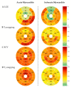
Location of LGE and Elevated Mapping Parameters per AHA Segments. Heatmapped 17-segment-model schemes (according to the American Heart Association) illustrate the percentage frequency of the occurrence of (A) LGE, (B) elevated T1, (C) elevated extracellular volume fraction (ECV) and (D) elevated T2.
Figure 4
Figure 4
Appearance of Acute and Subacute Myocarditis in CMR. (A) Acute myocarditis often demonstrates more obvious alterations of tissue characterization parameters including high prevalence and extent of late gadolinium enhancement (LGE) and elevated T2. (B) Subacute myocarditis can manifest with marked occurrence of LGE and elevated T1, ECV and T2; but in many cases may demonstrate rather subtle changes in tissue characterization.

References

    1. Imazio M., Trinchero R. Myopericarditis: Etiology, management, and prognosis. Int. J. Cardiol. 2008;127:17–26. doi: 10.1016/j.ijcard.2007.10.053. - DOI - PubMed
    1. Lampejo T., Durkin S.M., Bhatt N., Guttmann O. Acute myocarditis: Aetiology, diagnosis and management. Clin. Med. 2021;21:e505–e510. doi: 10.7861/clinmed.2021-0121. - DOI - PMC - PubMed
    1. Law Y.M., Lal A.K., Chen S., Čiháková D., Cooper L.T., Jr., Deshpande S., Godown J., Grosse-Wortmann L., Robinson J.D., Towbin J.A., et al. Diagnosis and Management of Myocarditis in Children. Circulation. 2021;144:e123–e125. doi: 10.1161/CIR.0000000000001001. - DOI - PubMed
    1. Ferreira V.M., Schulz-Menger J., Holmvang G., Kramer C.M., Carbone I., Sechtem U., Kindermann I., Gutberlet M., Cooper L.T., Liu P., et al. Cardiovascular Magnetic Resonance in Nonischemic Myocardial Inflammation: Expert Recommendations. J. Am. Coll. Cardiol. 2018;72:3158–3176. doi: 10.1016/j.jacc.2018.09.072. - DOI - PubMed
    1. Caforio A.L.P., Pankuweit S., Arbustini E., Basso C., Gimeno-Blanes J., Felix S.B., Fu M., Heliö T., Heymans S., Jahns R., et al. Current state of knowledge on aetiology, diagnosis, management, and therapy of myocarditis: A position statement of the European Society of Cardiology Working Group on Myocardial and Pericardial Diseases. Eur. Heart J. 2013;34:2636–2648. doi: 10.1093/eurheartj/eht210. - DOI - PubMed

LinkOut - more resources