

Risk Factors for Progression of Age-Related Macular Degeneration: Population-Based Amish Eye Study
- PMID: 36079043
- PMCID: PMC9457199
- DOI: 10.3390/jcm11175110
Risk Factors for Progression of Age-Related Macular Degeneration: Population-Based Amish Eye Study
Abstract
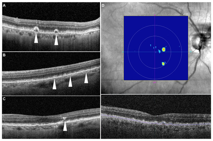
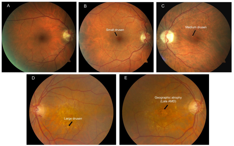
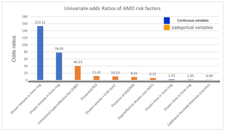
Objective: To evaluate the optical coherence tomography (OCT)-based risk factors for progression to late age-related macular degeneration (AMD) in a population-based study of elderly Amish. Methods: A total of 1332 eyes of 666 consecutive subjects who completed a 2-year follow-up visit were included in this multicenter, prospective, longitudinal, observational study. Imaging features were correlated with 2-year incidence of late AMD development. Odds ratios for imaging features were estimated from logistic regression. Baseline OCT images were reviewed for the presence of drusen volume ≥0.03 mm3 in the central 3 mm ring, intraretinal hyperreflective foci (IHRF), hyporeflective drusen cores (hDC), subretinal drusenoid deposits (SDD), and drusenoid pigment epithelium detachment (PED). Subfoveal choroidal thickness, drusen area, and drusen volume within 3 and 5 mm circles centered on the fovea were also assessed. Results: Twenty-one (1.5%) of 1332 eyes progressed to late AMD by 2 years. The mean age of the study subjects was 65 ± 10.17 (±SD) years and 410 subjects were female. Univariate logistic regression showed that drusen area and volume in both 3 mm and 5 mm circles, subfoveal choroidal thickness, drusen volume ≥ 0.03 mm3 in the 3 mm ring, SDD, IHRF, and hDC were all associated with an increased risk for development of late AMD. The multivariate regression model identified that drusen volume in the 3 mm ring (OR: 2.59, p = 0.049) and presence of IHRF (OR: 57.06, p < 0.001) remained as independent and significant risk factors for progression to late AMD. Conclusions: This population-based study confirms previous findings from clinic-based studies that high central drusen volume and IHRF are associated with an increased risk of progression to late AMD. These findings may be of value in risk-stratifying patients in clinical practice or identifying subjects for early intervention clinical trials.
Keywords: Amish eye study; age-related macular degeneration; complete retinal pigment epithelial and outer retina atrophy; geographic atrophy; optical coherence tomography.
Conflict of interest statement
M.G. Nittala, F. Corvi, J. Maram, S.B. Velaga, J.L. Haines, M.A. Pericak-Vance, D. Stambolian declare no conflicts of interest. S.R. Sadda reports consulting fees from Amgen, Allergan, Regeneron, Roche/Genentech, Novartis, Merck, 4DMT, Optos, Heidelberg, and Centervue. He also receives research instruments from Topcon, Nidek, Heidelberg, Centervue, Optos, and Carl Zeiss Meditec, outside the submitted work.
Figures
References
-
- Wong W.L., Su X., Li X., Cheung C.M.G., Klein R., Cheng C.-Y., Wong T.Y. Global Prevalence of Age-Related Macular Degeneration and Disease Burden Projection for 2020 and 2040: A Systematic Review and Meta-Analysis. Lancet Glob. Health. 2014;2:e106–e116. doi: 10.1016/S2214-109X(13)70145-1. - DOI - PubMed
-
- Age-Related Eye Disease Study Research Group Risk Factors Associated with Age-Related Macular Degeneration: A Case-Control Study in the Age-Related Eye Disease Study: Age-Related Eye Disease Study Report Number 3. Ophthalmology. 2000;107:2224–2232. doi: 10.1016/S0161-6420(00)00409-7. - DOI - PMC - PubMed
-
- Naj A.C., Scott W.K., Courtenay M.D., Cade W.H., Schwartz S.G., Kovach J.L., Agarwal A., Wang G., Haines J.L., Pericak-Vance M.A. Genetic Factors in Nonsmokers with Age-Related Macular Degeneration Revealed through Genome-Wide Gene-Environment Interaction Analysis. Ann. Hum. Genet. 2013;77:215–231. doi: 10.1111/ahg.12011. - DOI - PMC - PubMed
-
- Holz F.G., Sadda S.R., Busbee B., Chew E.Y., Mitchell P., Tufail A., Brittain C., Ferrara D., Gray S., Honigberg L., et al. Efficacy and Safety of Lampalizumab for Geographic Atrophy Due to Age-Related Macular Degeneration: Chroma and Spectri Phase 3 Randomized Clinical Trials. JAMA Ophthalmol. 2018;136:666–677. doi: 10.1001/jamaophthalmol.2018.1544. - DOI - PMC - PubMed
Grants and funding
LinkOut - more resources
Full Text Sources
