

Effects of Different Pressure Levels in Topical Negative Pressure Application-Analysis of Perfusion Parameters in a Clinical Skin Model Using Multimodal Imaging Techniques
- PMID: 36079063
- PMCID: PMC9457425
- DOI: 10.3390/jcm11175133
Effects of Different Pressure Levels in Topical Negative Pressure Application-Analysis of Perfusion Parameters in a Clinical Skin Model Using Multimodal Imaging Techniques
Abstract
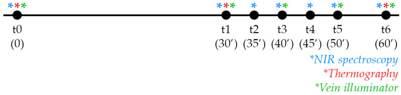
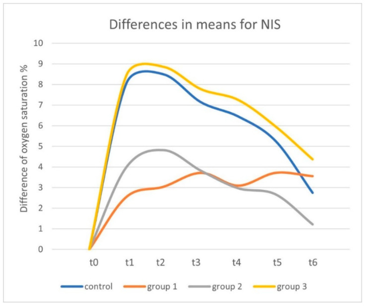
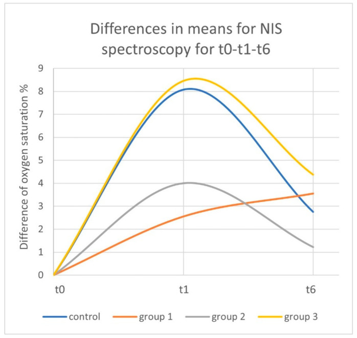
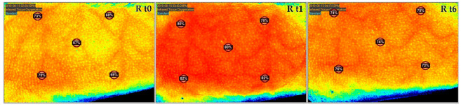
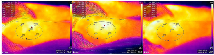
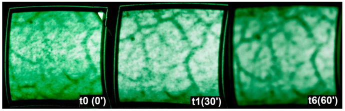
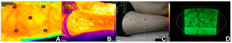
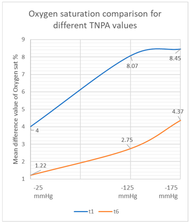
The effects of topical negative pressure therapy (TNP) have been a subject of research for many years. In this study, we investigated new imaging devices to detect clinical changes that TNP causes on healthy tissue and identified differences in microcirculation created by different pressure levels. We used near-infrared spectroscopy (NIS), thermography, and a vein illuminator to measure the differences in oxygen saturation, tissue temperature, and vein pattern. A control group (-125 mmHg) and three comparison groups with only TNP dressing (Group 1), -25 mmHg (Group 2), and -175 mmHg (Group 3) were established. Thirty minutes of TNP on intact skin was followed by 30 min of resting. A total of 24 participants were measured by all imaging devices at predetermined time points. Oxygen saturation and skin temperature increased by 8.07% and 1.67 °C for the control group, 4.00% and 1.65 °C for Group 2, and 8.45% and 1.68 °C for Group 3. Group 1 showed a slight increase in oxygen saturation and a 2.7 °C increase in skin temperature. Over the 30 min following removal of TNP, oxygen saturation and temperature decreased gradually for all groups. The vein illuminator did not show significant differences in the venous pattern or flow. Our study showed that higher negative pressure values resulted in higher oxygen saturation and higher tissue temperature.
Keywords: imaging; near infrared spectroscopy; negative pressure wound therapy; perfusion; pressure level; thermography.
Conflict of interest statement
R. E. Horch has received third-party funding for scientific research on NPWT from KCI—an Acelity company—in the past and has served as a member of a Scientific Advisory Board of KCI—Acelity in the past. R. E. Horch and A. Arkudas served as speakers on scientific symposia of KCI—Acelity in the past. The authors have no other relevant affiliations or financial involvement with any organization or entity with a financial interest in or financial conflict with the subject matter or materials discussed in the manuscript apart from those disclosed. The other authors declare no conflict of interest.
Figures

References
-
- Horch R.E., Gerngross H., Lang W., Mauckner P., Nord D., Peter R.U., Vogt P.M., Wetzel-Roth W., Willy C. Indications and safety aspects of vacuum-assisted wound closure. MMW Fortschr. Med. 2005;147((Suppl. S1)):1–5. - PubMed
-
- Horch R.E., Braumann C., Dissemond J., Lehner B., Hirche C., Woeste G., Wetzel-Roth W., Willy C. Use of Negative Pressure Wound Therapy with Instillation and Dwell Time for Wound Treatment—Results of an Expert Consensus Conference. Zentralblatt Chir. 2018;143:609–616. doi: 10.1055/a-0713-0517. - DOI - PubMed
LinkOut - more resources
Full Text Sources
