. 2022 Aug 28;15(17):5947.
doi: 10.3390/ma15175947.

Evaluation of Incipient Enamel Caries at Smooth Tooth Surfaces Using SS-OCT

Affiliations

Evaluation of Incipient Enamel Caries at Smooth Tooth Surfaces Using SS-OCT

Yasushi Shimada et al. Materials (Basel). .

Abstract

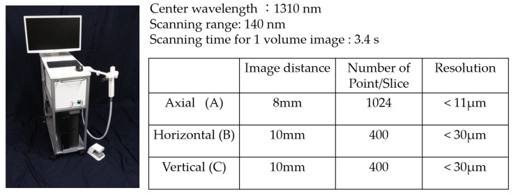
(1) Background: Dental caries, if diagnosed at the initial stage, can be arrested and remineralized by a non-operative therapeutic approach preserving tooth structure. Accurate and reproducible diagnostic procedure is required for the successful management of incipient caries. The aim of this study was to evaluate the diagnostic accuracy of 3D swept-source optical coherence tomography (3D SS-OCT) for enamel caries at smooth tooth surface if the lesion was with remineralization. (2) Methods: Forty-seven tooth surfaces of 24 extracted human teeth visibly with/without enamel caries (ICDAS code 0−3) were selected and used in this study. The tooth surfaces of investigation site were cleaned and visually examined by four dentists. After the visual inspection, SS-OCT scanning was performed onto the enamel surfaces to construct a 3D image. The 2D tomographic images of the investigation site were chosen from the 3D dataset and dynamically displayed in video and evaluated by the examiners. A five-rank scale was used to score the level of enamel caries according to the following; 1: Intact enamel. 2: Noncavitated lesion with remineralization. 3: Superficial noncavitated lesion without remineralization. 4: Deep nonvacitated lesion without remineralization. 5: Enamel lesion with cavitation. Sensitivity and specificity for 3D OCT image and visual inspection were calculated. Diagnostic accuracy of each diagnostic method was calculated using weighted kappa. Statistical significance was defined at p = 0.05. (3) Results: 3D SS-OCT could clearly depict enamel caries at smooth tooth surface as a bright zone, based on the increased backscattering signal. It was noted that 3D SS-OCT showed higher sensitivity for the diagnosis of remineralized lesions and deep enamel lesions without cavitation, as well as cavitated enamel lesions (p < 0.05). No significant difference of specificity was observed between the two diagnostic methods (p > 0.05). Furthermore, 3D SS-OCT showed higher diagnostic accuracy than visual inspection (p < 0.05). (4) Conclusions: Within the limitations of this in vitro study, 3D SS-OCT showed higher diagnostic capacity for smooth surface enamel caries than visual inspection and could also discriminate lesion remineralization of enamel caries.

Keywords: 3D image; caries; demineralization; diagnosis; enamel; optical coherence tomography; remineralization.


Conflict of interest statement

The authors declare no conflict of interest.

Figures

Figure 1
Figure 1
SS-OCT system used (Prototype Yoshida Dental OCT).
Figure 2
Figure 2
Images of deep enamel demineralization (score 2). (a) Front view of enamel demineralization; (b) Histological view of the enamel demineralization sectioned horizontally; (c) CLSM image. Morphological change within the demineralization is evident. Enamel demineralization penetrates inner half of enamel (white arrow).
Figure 3
Figure 3
3D CLSM images of deep enamel demineralization (score 2). 3D image of Figure 2c. Zone of demineralization is clearly imaged as loss of surface (white arrow).
Figure 4
Figure 4
SS-OCT Images of deep enamel demineralization (score 2). Same tooth as Figure 2. (a) 3D SS-OCT image. Due to the scattering from the internal structure, enamel demineralization is clearly shown as a white zone (arrow); (b) 2D SS-OCT image chosen from the 3D image (a) along red line. Demineralization penetrates enamel inner zone (arrow). Dynamic slicing 3D video is presented in Supplementary Materials Video S1.
Figure 5
Figure 5
Images of remineralized enamel demineralization (score 3). (a) Front view of enamel demineralization; (b) Histological view of enamel lesion sectioned horizontally; (c) CLSM image. Morphological change within the lesion is not so evident (black arrow). Surface enamel of the lesion morphologically continues with surrounding enamel, suggesting the remineralization (white arrow).
Figure 6
Figure 6
3D CLSM images of remineralized enamel demineralization (score 3). 3D image of Figure 5c. Zone of demineralization is not evident (white arrow).
Figure 7
Figure 7
SS-OCT images of remineralized enamel demineralization (score 3). Same tooth as Figure 3. (a) 3D SS-OCT image of remineralized enamel demineralization. Due to the scattering from the subsurface lesion, presence of demineralization is imaged (arrow); (b) 2D SS-OCT image chosen from the 3D image (a) along red line. Formation of remineralization zone of lesion surface can be observed (arrow). Dynamic slicing 3D video is presented in Supplementary Materials Video S2.

Similar articles

Cited by

References

    1. Pitts N.B., Zero D.T., Marsh P.D., Ekstrand K., Weintraub J.A., Ramos-Gomez F., Tagami J., Twetman S., Tsakos G., Ismail A. Dental caries. Nat. Rev. Dis. Primers. 2017;3:17030. doi: 10.1038/nrdp.2017.30. - DOI - PubMed
    1. Featherstone J.D.B. The Continuum of Dental Caries—Evidence for a Dynamic Disease Process. J. Dent. Res. 2004;83((Suppl. 1)):C39–C42. doi: 10.1177/154405910408301s08. - DOI - PubMed
    1. Amaechi B.T. Remineralisation—The buzzword for early MI caries management. Br. Dent. J. 2017;223:173–182. doi: 10.1038/sj.bdj.2017.663. - DOI - PubMed
    1. Paris S., Meyer-Lueckel H., Kielbassa A. Resin Infiltration of Natural Caries Lesions. J. Dent. Res. 2007;86:662–666. doi: 10.1177/154405910708600715. - DOI - PubMed
    1. Meyer-Lueckel H., Wardius A., Krois J., Bitter K., Moser C., Paris S., Wierichs R. Proximal Caries Infiltration—Pragmatic RCT with 4 years of follow-up. J. Dent. 2021;111:103733. doi: 10.1016/j.jdent.2021.103733. - DOI - PubMed

LinkOut - more resources