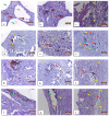
Long-Term Examination of Degradation and In Vivo Biocompatibility of Some Mg-0.5Ca-xY Alloys in Sprague Dawley Rats
- PMID: 36079340
- PMCID: PMC9456631
- DOI: 10.3390/ma15175958
Long-Term Examination of Degradation and In Vivo Biocompatibility of Some Mg-0.5Ca-xY Alloys in Sprague Dawley Rats
Abstract
The medical field has undergone constant development in recent years, and a segment of this development is occupied by biodegradable alloys. The most common alloys in this field are those based on Mg, their main advantage being the ability to degrade gradually, without affecting the patient, and also their ability to be fully absorbed by the human body. One of their most important conditions is the regeneration and replacement of human tissue. Tissue can be engineered in different ways, one being tissue regeneration in vivo, which can serve as a template. In vivo remodeling aims to restore tissue or organs. The key processes of tissue formation and maturation are: proliferation (sorting and differentiation of cells), proliferation and organization of the extracellular matrix, biodegradation of the scaffold-remodeling, and potential tissue growth. In the present paper, the design of the alloys in the Mg-Ca-Y system is formed from the beginning using high-purity components, Mg-98.5%, master-alloys: Mg-Y (70 wt.%-30 wt.%) and Mg-Ca (85 wt.%-15 wt.%). After 8 weeks of implantation, the degradation of the implanted material is observed, and only small remaining fragments are found. At the site of implantation, no inflammatory reaction is observed, but it is observed that the process of integration and reabsorption, over time, accentuates the prosaic surface of the material. The aim of the work is to test the biocompatibility of magnesium-based alloys on laboratory rats in order to use these alloys in medical applications. The innovative parts of these analyses are the chemical composition of the alloys used and the tests performed on laboratory animals.
Keywords: Mg-Ca-Y biodegradable alloys; in vivo animal study; surface morphology.
Conflict of interest statement
The authors declare no conflict of interest.
Figures


Similar articles
-
In vitro and in vivo assessment of biomedical Mg-Ca alloys for bone implant applications.J Appl Biomater Funct Mater. 2018 Jul;16(3):126-136. doi: 10.1177/2280800017750359. Epub 2018 Apr 2. J Appl Biomater Funct Mater. 2018. PMID: 29607729
-
In Vitro and In Vivo Analysis of the Mg-Ca-Zn Biodegradable Alloys.J Funct Biomater. 2024 Jun 17;15(6):166. doi: 10.3390/jfb15060166. J Funct Biomater. 2024. PMID: 38921539 Free PMC article.
-
Electrochemical Analysis and In Vitro Assay of Mg-0.5Ca-xY Biodegradable Alloys.Materials (Basel). 2020 Jul 10;13(14):3082. doi: 10.3390/ma13143082. Materials (Basel). 2020. PMID: 32664267 Free PMC article.
-
Design of magnesium alloys with controllable degradation for biomedical implants: From bulk to surface.Acta Biomater. 2016 Nov;45:2-30. doi: 10.1016/j.actbio.2016.09.005. Epub 2016 Sep 6. Acta Biomater. 2016. PMID: 27612959 Review.
-
Opportunities and challenges for the biodegradable magnesium alloys as next-generation biomaterials.Regen Biomater. 2016 Jun;3(2):79-86. doi: 10.1093/rb/rbw003. Epub 2016 Mar 23. Regen Biomater. 2016. PMID: 27047673 Free PMC article. Review.
Cited by
-
A Novel PLLA/MgF2 Coating on Mg Alloy by Ultrasonic Atomization Spraying for Controlling Degradation and Improving Biocompatibility.Materials (Basel). 2023 Jan 10;16(2):682. doi: 10.3390/ma16020682. Materials (Basel). 2023. PMID: 36676415 Free PMC article.
-
Challenges and Pitfalls of Research Designs Involving Magnesium-Based Biomaterials: An Overview.Int J Mol Sci. 2024 Jun 5;25(11):6242. doi: 10.3390/ijms25116242. Int J Mol Sci. 2024. PMID: 38892430 Free PMC article. Review.
References
-
- Bița A.I., Antoniac I., Miculescu M., Stan G.E., Leonat L., Antoniac A., Constantin B., Forna N. Electrochemical and In Vitro Biological Evaluation of Bio-Active Coatings Deposited by Magnetron Sputtering onto Biocompatible Mg-0.8Ca Alloy. Materials. 2022;15:3100. doi: 10.3390/ma15093100. - DOI - PMC - PubMed
-
- Arcos D., Vallet-Regí M. Bioceramics for drug delivery. Acta Mater. 2013;61:890–911. doi: 10.1016/j.actamat.2012.10.039. - DOI
LinkOut - more resources
Full Text Sources
Miscellaneous

