Manufacturing of Zinc Oxide Nanoparticle (ZnO NP)-Loaded Polyvinyl Alcohol (PVA) Nanostructured Mats Using Ginger Extract for Tissue Engineering Applications
- PMID: 36080077
- PMCID: PMC9457793
- DOI: 10.3390/nano12173040
Manufacturing of Zinc Oxide Nanoparticle (ZnO NP)-Loaded Polyvinyl Alcohol (PVA) Nanostructured Mats Using Ginger Extract for Tissue Engineering Applications
Abstract
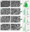
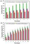
In this research, as an alternative to chemical and physical methods, environmentally and cost-effective antimicrobial zinc oxide nanoparticles (ZnO NP) were produced by the green synthesis method. The current study focuses on the production of ZnO NP starting from adequate precursor and Zingiber officinale aqueous root extracts (ginger). The produced ZnO NP was loaded into electrospun nanofibers at different concentrations for various tissue engineering applications such as wound dressings. The produced ZnO NPs and ZnO NP-loaded nanofibers were examined by Scanning Electron Microscopy (SEM) for morphological assessments and Fourier-transform infrared spectrum (FT-IR) for chemical assessments. The disc diffusion method was used to test the antimicrobial activity of ZnO NP and ZnO NP-loaded nanofibers against three representatives strains, Escherichia coli (Gram-negative bacteria), Staphylococcus aureus (Gram-positive bacteria), and Candida albicans (fungi) microorganisms. The strength and stretching of the produced fibers were assessed using tensile tests. Since water absorption and weight loss behaviors are very important in tissue engineering applications, swelling and degradation analyses were applied to the produced nanofibers. Finally, the MTT test was applied to analyze biocompatibility. According to the findings, ZnO NP-loaded nanofibers were successfully synthesized using a green precipitation approach and can be employed in tissue engineering applications such as wound dressing.
Keywords: ZnO NPs; antimicrobial effect; electrospinning; tissue engineering; wound dressing.
Conflict of interest statement
The authors declare that they have no financial interest in this paper.
Figures


References
-
- Danie Kingsley J., Ranjan S., Dasgupta N., Saha P. Nanotechnology for tissue engineering: Need, techniques and applications. J. Pharm. Res. 2013;7:200–204. doi: 10.1016/j.jopr.2013.02.021. - DOI
-
- Ilhan E., Ulag S., Sahin A., Ekren N., Kilic O., Oktar F.N., Gunduz O. Lecture Notes in Computer Science (İncluding Subseries Lecture Notes in Artificial Intelligence and Lecture Notes in Bioinformatics) Springer; Cham, Switzerland: 2020. Production of 3D-printed tympanic membrane scaffolds as a tissue engineering application.
-
- Tasci M.E., Dede B., Tabak E., Gur A., Sulutas R.B., Cesur S., Ilhan E., Lin C.C., Paik P., Ficai D., et al. Production, optimization and characterization of polylactic acid microparticles using electrospray with porous structure. Appl. Sci. 2021;11:5090. doi: 10.3390/app11115090. - DOI
-
- Geltmeyer J., Van der Schueren L., Goethals F., De Buysser K., De Clerck K. Optimum sol viscosity for stable electrospinning of silica nanofibres. J. Sol-Gel Sci. Technol. 2013;67:188–195. doi: 10.1007/s10971-013-3066-x. - DOI
LinkOut - more resources
Full Text Sources
Miscellaneous

