

Current Research on Zinc Oxide Nanoparticles: Synthesis, Characterization, and Biomedical Applications
- PMID: 36080103
- PMCID: PMC9459703
- DOI: 10.3390/nano12173066
Current Research on Zinc Oxide Nanoparticles: Synthesis, Characterization, and Biomedical Applications
Abstract
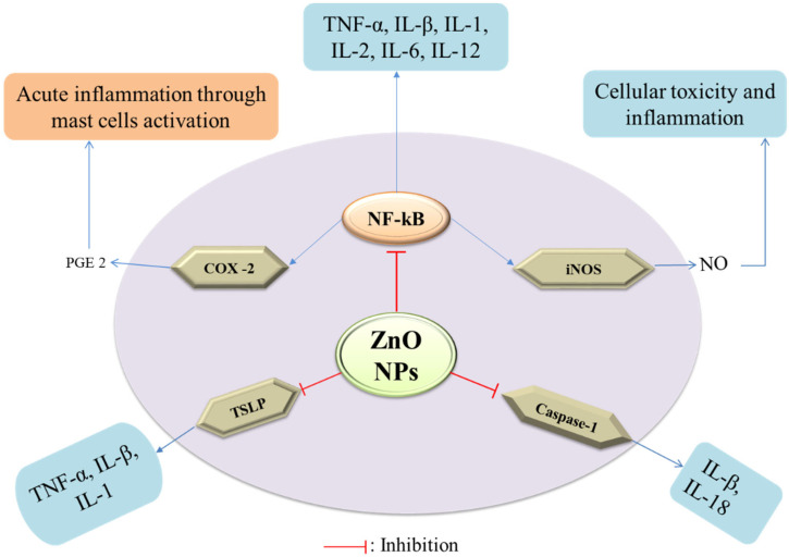
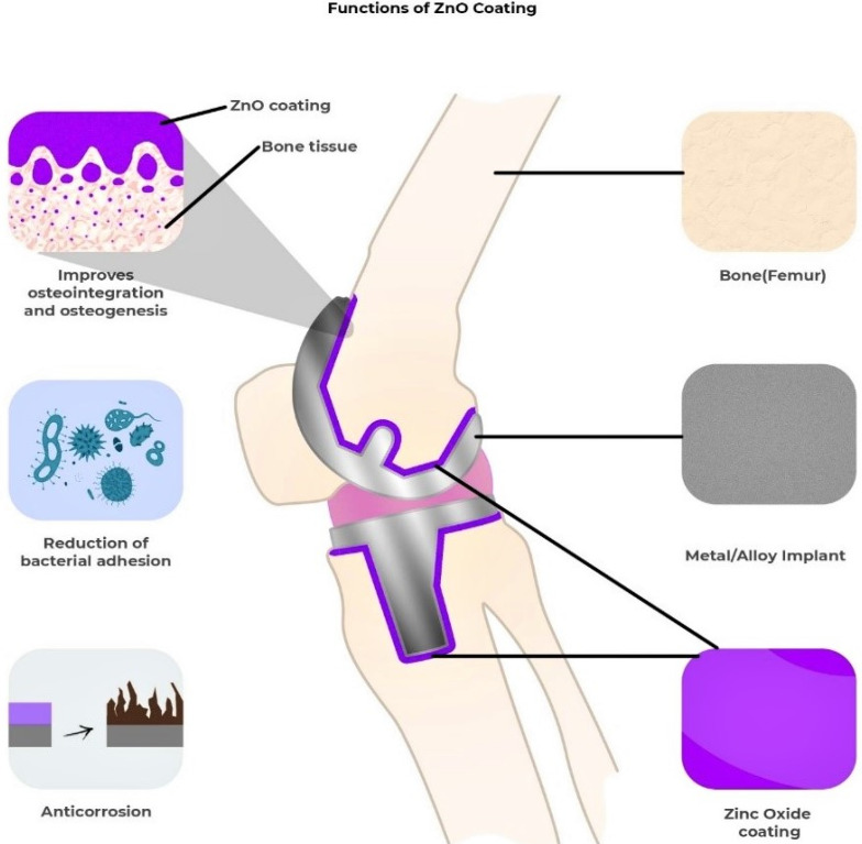
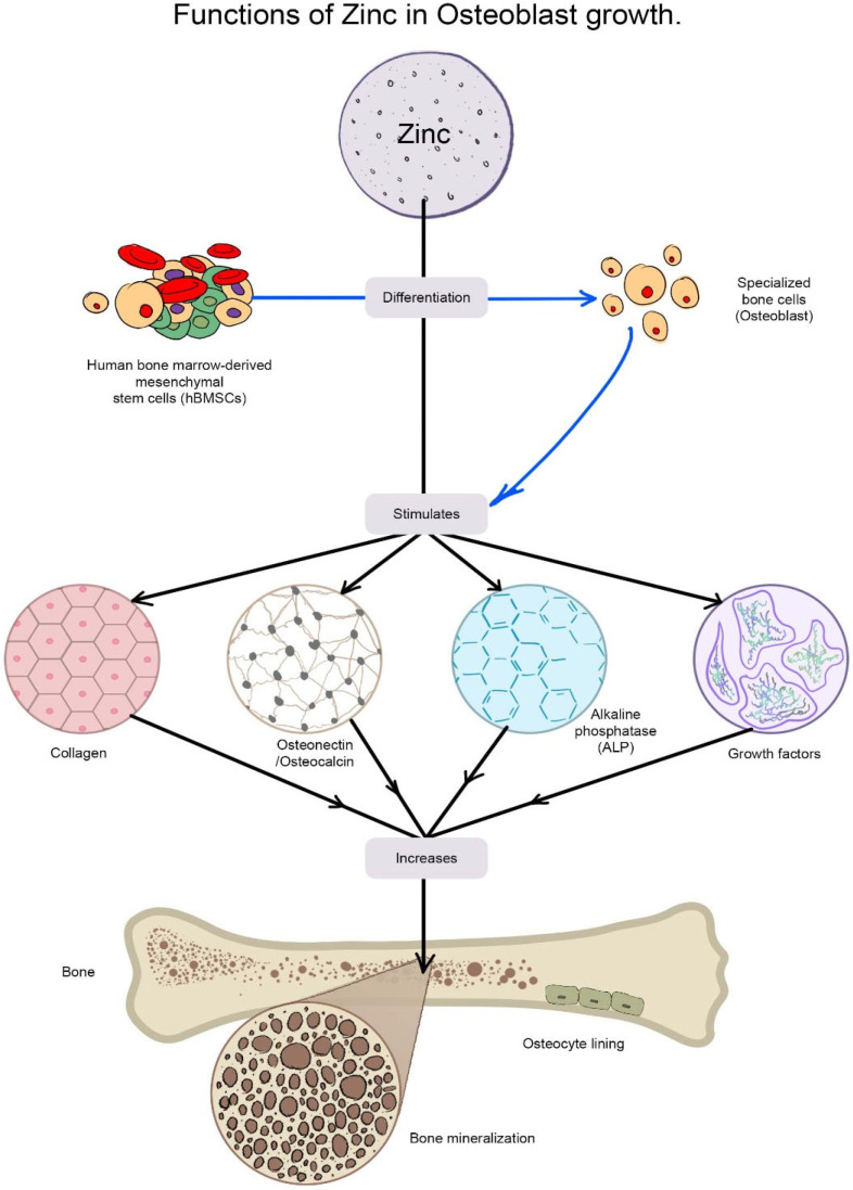
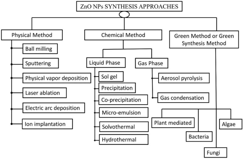
Zinc oxide nanoparticles (ZnO-NPs) have piqued the curiosity of researchers all over the world due to their extensive biological activity. They are less toxic and biodegradable with the capacity to greatly boost pharmacophore bioactivity. ZnO-NPs are the most extensively used metal oxide nanoparticles in electronic and optoelectronics because of their distinctive optical and chemical properties which can be readily modified by altering the morphology and the wide bandgap. The biosynthesis of nanoparticles using extracts of therapeutic plants, fungi, bacteria, algae, etc., improves their stability and biocompatibility in many biological settings, and its biofabrication alters its physiochemical behavior, contributing to biological potency. As such, ZnO-NPs can be used as an effective nanocarrier for conventional drugs due to their cost-effectiveness and benefits of being biodegradable and biocompatible. This article covers a comprehensive review of different synthesis approaches of ZnO-NPs including physical, chemical, biochemical, and green synthesis techniques, and also emphasizes their biopotency through antibacterial, antifungal, anticancer, anti-inflammatory, antidiabetic, antioxidant, antiviral, wound healing, and cardioprotective activity. Green synthesis from plants, bacteria, and fungus is given special attention, with a particular emphasis on extraction techniques, precursors used for the synthesis and reaction conditions, characterization techniques, and surface morphology of the particles.
Keywords: biological activities; green synthesis; zinc oxide nanoparticles.
Conflict of interest statement
The authors declare no conflict of interest.
Figures
Similar articles
-
Recent advances in the synthesis, characterization and biomedical applications of zinc oxide nanoparticles.Bioprocess Biosyst Eng. 2023 Oct;46(10):1377-1398. doi: 10.1007/s00449-023-02886-1. Epub 2023 Jun 9. Bioprocess Biosyst Eng. 2023. PMID: 37294320 Free PMC article. Review.
-
A Review of the Green Synthesis of ZnO Nanoparticles Utilising Southern African Indigenous Medicinal Plants.Nanomaterials (Basel). 2022 Oct 3;12(19):3456. doi: 10.3390/nano12193456. Nanomaterials (Basel). 2022. PMID: 36234584 Free PMC article. Review.
-
Biofabrication and characterization of cyanobacteria derived ZnO NPs for their bioactivity comparison with commercial chemically synthesized nanoparticles.Bioorg Chem. 2021 Aug;113:104999. doi: 10.1016/j.bioorg.2021.104999. Epub 2021 May 21. Bioorg Chem. 2021. PMID: 34062406
-
Antibacterial and Photodegradation of Organic Dyes Using Lamiaceae-Mediated ZnO Nanoparticles: A Review.Nanomaterials (Basel). 2022 Dec 16;12(24):4469. doi: 10.3390/nano12244469. Nanomaterials (Basel). 2022. PMID: 36558321 Free PMC article. Review.
-
Desertifilum sp. EAZ03 cell extract as a novel natural source for the biosynthesis of zinc oxide nanoparticles and antibacterial, anticancer and antibiofilm characteristics of synthesized zinc oxide nanoparticles.J Appl Microbiol. 2022 Jan;132(1):221-236. doi: 10.1111/jam.15177. Epub 2021 Jul 19. J Appl Microbiol. 2022. PMID: 34101961
Cited by
-
Anticancer effect of zinc oxide nanoparticles prepared by varying entry time of ion carriers against A431 skin cancer cells in vitro.Front Chem. 2022 Dec 1;10:1069450. doi: 10.3389/fchem.2022.1069450. eCollection 2022. Front Chem. 2022. PMID: 36531331 Free PMC article.
-
Nanoparticle‑based antiviral strategies to combat the influenza virus (Review).Biomed Rep. 2024 Feb 21;20(4):65. doi: 10.3892/br.2024.1753. eCollection 2024 Apr. Biomed Rep. 2024. PMID: 38476608 Free PMC article. Review.
-
Recycled or Bio-Based Solvents for the Synthesis of ZnO Nanoparticles: Characterization and Validation in Organic Solar Cells.Materials (Basel). 2024 Mar 14;17(6):1332. doi: 10.3390/ma17061332. Materials (Basel). 2024. PMID: 38541486 Free PMC article.
-
Dietary Transfer of Zinc Oxide Nanoparticles Induces Locomotive Defects Associated with GABAergic Motor Neuron Damage in Caenorhabditis elegans.Nanomaterials (Basel). 2023 Jan 10;13(2):289. doi: 10.3390/nano13020289. Nanomaterials (Basel). 2023. PMID: 36678041 Free PMC article.
-
Study of the effect of zinc oxide, selenium, and silver nanoparticles on the expression level of oxidative stress-associated genes in ovarian cancer.Med Oncol. 2025 Jan 6;42(2):39. doi: 10.1007/s12032-024-02593-1. Med Oncol. 2025. PMID: 39760958
References
-
- Agarwal H., Kumar S.V., Rajeshkumar S. A review on green synthesis of zinc oxide nanoparticles—An eco-friendly approach. Resour. Technol. 2017;3:406–413. doi: 10.1016/j.reffit.2017.03.002. - DOI
-
- Rodnyi P.A., Khodyuk I.V. Optical and luminescence properties of zinc oxide (Review) Opt. Spectrosc. 2011;111:776–785. doi: 10.1134/S0030400X11120216. - DOI
-
- Shaba E.Y., Jacob J.O., Tijani J.O., Suleiman M.A.T. A critical review of synthesis parameters affecting the properties of zinc oxide nanoparticle and its application in wastewater treatment. Appl. Water Sci. 2021;11:48. doi: 10.1007/s13201-021-01370-z. - DOI
-
- Kielbik P., Kaszewski J., Rosowska J., Wolska E., Witkowski B., Gralak M., Gajewski Z., Godlewski M., Godlewski M.M. Biodegradation of the ZnO:Eu nanoparticles in the tissues of adult mouse after alimentary application. Nanomed. Nanotechnol. Biol. Med. 2017;13:843–852. doi: 10.1016/j.nano.2016.11.002. - DOI - PubMed
Publication types
LinkOut - more resources
Full Text Sources
Other Literature Sources
Research Materials
