

Degradative Effect of Nattokinase on Spike Protein of SARS-CoV-2
- PMID: 36080170
- PMCID: PMC9458005
- DOI: 10.3390/molecules27175405
Degradative Effect of Nattokinase on Spike Protein of SARS-CoV-2
Abstract
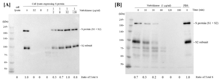
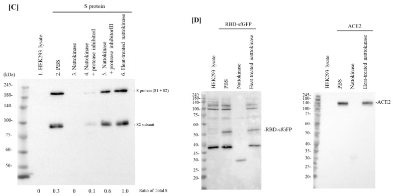
The coronavirus disease 2019 (COVID-19), caused by the severe acute respiratory syndrome coronavirus 2 (SARS-CoV-2), emerged as a pandemic and has inflicted enormous damage on the lives of the people and economy of many countries worldwide. However, therapeutic agents against SARS-CoV-2 remain unclear. SARS-CoV-2 has a spike protein (S protein), and cleavage of the S protein is essential for viral entry. Nattokinase is produced by Bacillus subtilis var. natto and is beneficial to human health. In this study, we examined the effect of nattokinase on the S protein of SARS-CoV-2. When cell lysates transfected with S protein were incubated with nattokinase, the S protein was degraded in a dose- and time-dependent manner. Immunofluorescence analysis showed that S protein on the cell surface was degraded when nattokinase was added to the culture medium. Thus, our findings suggest that nattokinase exhibits potential for the inhibition of SARS-CoV-2 infection via S protein degradation.
Keywords: COVID-19; SARS-CoV-2; nattokinase.
Conflict of interest statement
The authors declare no conflict of interest.
Figures
Similar articles
-
The TMPRSS2 Inhibitor Nafamostat Reduces SARS-CoV-2 Pulmonary Infection in Mouse Models of COVID-19.mBio. 2021 Aug 31;12(4):e0097021. doi: 10.1128/mBio.00970-21. Epub 2021 Aug 3. mBio. 2021. PMID: 34340553 Free PMC article.
-
Inhibitory effect of honokiol on furin-like activity and SARS-CoV-2 infection.J Tradit Complement Med. 2022 Jan;12(1):69-72. doi: 10.1016/j.jtcme.2021.09.005. Epub 2021 Sep 16. J Tradit Complement Med. 2022. PMID: 34545325 Free PMC article.
-
SARS-CoV-2 Spike Furin Cleavage Site and S2' Basic Residues Modulate the Entry Process in a Host Cell-Dependent Manner.J Virol. 2022 Jul 13;96(13):e0047422. doi: 10.1128/jvi.00474-22. Epub 2022 Jun 9. J Virol. 2022. PMID: 35678602 Free PMC article.
-
Microalgae as an Efficient Vehicle for the Production and Targeted Delivery of Therapeutic Glycoproteins against SARS-CoV-2 Variants.Mar Drugs. 2022 Oct 23;20(11):657. doi: 10.3390/md20110657. Mar Drugs. 2022. PMID: 36354980 Free PMC article. Review.
-
The expression of hACE2 receptor protein and its involvement in SARS-CoV-2 entry, pathogenesis, and its application as potential therapeutic target.Tumour Biol. 2021;43(1):177-196. doi: 10.3233/TUB-200084. Tumour Biol. 2021. PMID: 34420993 Review.
Cited by
-
Comparative Cardioprotective Effectiveness: NOACs vs. Nattokinase-Bridging Basic Research to Clinical Findings.Biomolecules. 2024 Aug 7;14(8):956. doi: 10.3390/biom14080956. Biomolecules. 2024. PMID: 39199344 Free PMC article. Review.
-
Novel Oronasal Drainage for Long COVID: Proposed Mechanisms-Case Report.Viruses. 2025 Jan 31;17(2):210. doi: 10.3390/v17020210. Viruses. 2025. PMID: 40006965 Free PMC article.
-
Clinical Approach to Post-acute Sequelae After COVID-19 Infection and Vaccination.Cureus. 2023 Nov 21;15(11):e49204. doi: 10.7759/cureus.49204. eCollection 2023 Nov. Cureus. 2023. PMID: 38024037 Free PMC article. Review.
-
Ongoing Treatment with a Spore-Based Probiotic Containing Five Strains of Bacillus Improves Outcomes of Mild COVID-19.Nutrients. 2023 Jan 17;15(3):488. doi: 10.3390/nu15030488. Nutrients. 2023. PMID: 36771194 Free PMC article.
-
Strategies for the Management of Spike Protein-Related Pathology.Microorganisms. 2023 May 17;11(5):1308. doi: 10.3390/microorganisms11051308. Microorganisms. 2023. PMID: 37317282 Free PMC article. Review.
References
-
- Bestle D., Heindl M.R., Limburg H., Van Lam van T., Pilgram O., Moulton H., Stein D.A., Hardes K., Eickmann M., Dolnik O., et al. TMPRSS2 and furin are both essential for proteolytic activation of SARS-CoV-2 in human airway cells. Life Sci. Alliance. 2020;3:e202000786. doi: 10.26508/lsa.202000786. - DOI - PMC - PubMed
MeSH terms
Substances
LinkOut - more resources
Full Text Sources
Miscellaneous
