. 2022 Aug 30;27(17):5574.
doi: 10.3390/molecules27175574.

Crystal Structure of Human CD47 in Complex with Engineered SIRPα.D1(N80A)

Affiliations

Crystal Structure of Human CD47 in Complex with Engineered SIRPα.D1(N80A)

Jifeng Yu et al. Molecules. .

Abstract

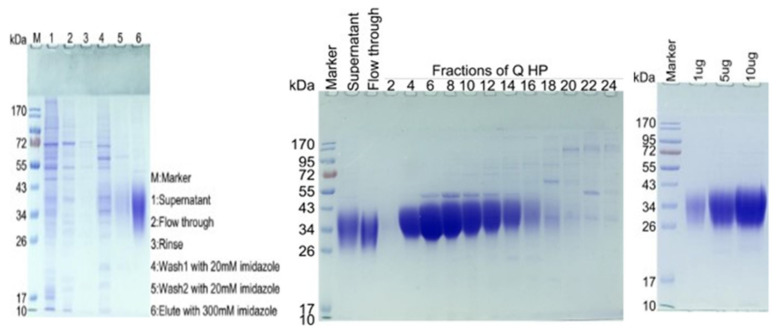
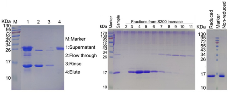
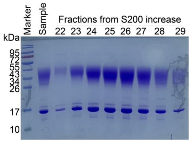
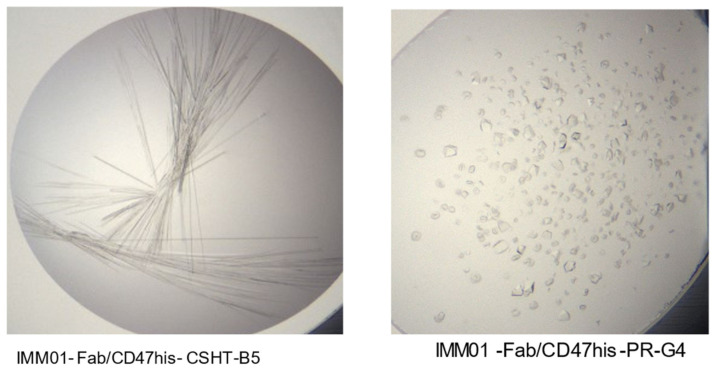
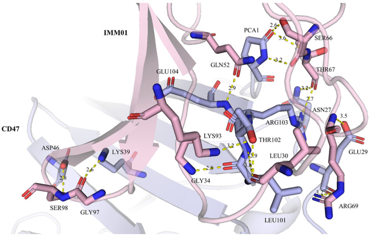
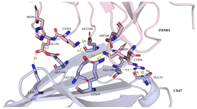
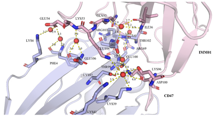
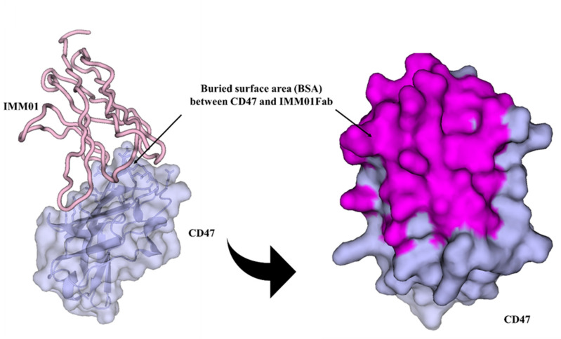
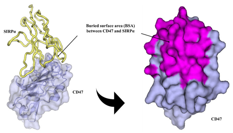
Background: Targeting the CD47/SIRPα signaling pathway represents a novel approach to enhance anti-tumor immunity. However, the crystal structure of the CD47/SIRPα has not been fully studied. This study aims to analyze the structure interface of the complex of CD47 and IMM01, a novel recombinant SIRPα-Fc fusion protein. Methods: IMM01-Fab/CD47 complex was crystalized, and diffraction images were collected. The complex structure was determined by molecular replacement using the program PHASER with the CD47-SIRPαv2 structure (PDB code 2JJT) as a search model. The model was manually built using the COOT program and refined using TLS parameters in REFMAC from the CCP4 program suite. Results: Crystallization and structure determination analysis of the interface of IMM01/CD47 structure demonstrated CD47 surface buried by IMM01. Comparison with the literature structure (PDB ID 2JJT) showed that the interactions of IMM01/CD47 structure are the same. All the hydrogen bonds that appear in the literature structure are also present in the IMM01/CD47 structure. These common hydrogen bonds are stable under different crystal packing styles, suggesting that these hydrogen bonds are important for protein binding. In the structure of human CD47 in complex with human SIRPα, except SER66, the amino acids that form hydrogen bonds are all conserved. Furthermore, comparing with the structure of PDB ID 2JJT, the salt bridge interaction from IMM01/CD47 structure are very similar, except the salt bridge bond between LYS53 in IMM01 and GLU106 in CD47, which only occurs between the B and D chains. However, as the side chain conformation of LYS53 in chain A is slightly different, the salt bridge bond is absent between the A and C chains. At this site between chain A and chain C, there are a salt bridge bond between LYS53 (A) and GLU104 (C) and a salt bridge bond between HIS56 (A) and GLU106 (C) instead. According to the sequence alignment results of SIRPα, SIRPβ and SIRPγ in the literature of PDB ID 2JJT, except ASP100, the amino acids that form common salt bridge bonds are all conserved. Conclusion: Our data demonstrated crystal structure of the IMM01/CD47 complex and provides a structural basis for the structural binding interface and future clinical applications.

Keywords: CD47/SIRPα; SIRPα-Fc fusion proteins; cancer immunotherapy; computer-aided drug discovery; crystal structure; immune checkpoint pathway.


Conflict of interest statement

Song Li, Dianze Chen, Dandan Liu, Huiqin Guo, Chunmei Yang, Wei Zhang, Li Zhang, Gui Zhao, Xiaoping Tu, Liang Peng, Sijin Liu, Xing Bai and Ruliang Zhang are employees of ImmuneOnco Biopharmaceuticals (Shanghai) Co., Ltd. Wenzhi Tian is the founder of ImmuneOnco Biopharmaceuticals (Shanghai) Co., Ltd. Jifeng Yu, Yongping Song and Zhongxing Jiang declare no conflicts of interest.

Figures

Figure 1
Figure 1
SDS-PAGE analysis of CD47-His after the Ni column showed a molecular weight of ~34 kDa (left figure). Based on the result, fractions of flow through 4–14 were pooled together and concentrated by an ultrafiltration tube with a molecular cut-off value of 10 kDa (middle figure). The SDS-PAGE analysis of final CD47-His showed a clean band with a molecular weight of ~34 kDa (right figure). The theoretical molecular weight of CD47-His is 14.8 kDa. The SDS-PAGE re-sults show that the actual molecular weight is ~34 kDa due to the severe glycosylation modifica-tion of CD47.
Figure 2
Figure 2
Protein A purification method was used to purify the IMM01-Fab from the supernatant (left figure). Superdex200 column was used increase purity of IMM01-Fab. Fractions 3, 4, 5 were pooled together for concentration (middle figure). SDS-PAGE analysis of final purified IMM01-Fab showed a clean single band (right figure).
Figure 3
Figure 3
IMM01-Fab and CD47-His were mixed in molar ratio 1:1 and incubated at 4 °C overnight. The size-exclusion chromatography (SEC) method was used for IMM01-Fab/CD47 complex purification. Superdex200 column was used to increase purity of IMM01-Fab/CD47 complex. Fractions 23–26 were concentrated for crystallization.
Figure 4
Figure 4
Crystal observation in two different conditions, IMM01-Fab/CD47his-CSHT-B5 (left figure) and IMM01-Fab/CD47his-PR-G4 (right figure).
Figure 5
Figure 5
Hydrogen bond interactions between chain A (IMM01) and chain C (CD47). Chain A and chain C are shown in pink and light blue, respectively. The residues forming hydrogen bonds are shown as sticks, key hydrogen bonds are highlighted as yellow dashed lines.
Figure 6
Figure 6
Salt bridge bond interactions between chain A (IMM01) and chain C (CD47). Chain A and chain C are shown in pink and light blue, respectively. The residues forming salt bridge bonds are shown as sticks, salt bridge bonds are highlighted as yellow dashed lines.
Figure 7
Figure 7
Water-bridged hydrogen bond interactions between chain A (IMM01) and chain C (CD47). Chain A and chain C are shown in pink and light blue, respectively. The residues forming hydrogen bond with waters are shown as sticks, and the water molecules are shown as red spheres. Key hydrogen bonds are highlighted as yellow dashed lines.
Figure 8
Figure 8
Buried surface area (BSA) between chain A (IMM01) and chain C (CD47). Chain A and chain C are shown in pink and light blue, respectively. The CD47 is shown as surface, and the interfacing residues are highlighted in magenta.
Figure 9
Figure 9
Buried surface area (BSA) between chain A (SIRPα, 2JJS) and chain C (CD47, 2JJS). Chain A and chain C are shown in yellow and light blue, respectively. The CD47 is shown as surface, and the interfacing residues are highlighted in magenta.

Similar articles

Cited by

References

    1. Jiang Z., Sun H., Yu J., Tian W., Song Y. Targeting CD47 for cancer immunotherapy. J. Hematol. Oncol. 2021;14:180. doi: 10.1186/s13045-021-01197-w. - DOI - PMC - PubMed
    1. Wang Y., Zhao C., Liu Y., Wang C., Jiang H., Hu Y., Wu J. Recent Advances of Tumor Therapy Based on the CD47-SIRPα Axis. Mol. Pharm. 2022;19:1273–1293. doi: 10.1021/acs.molpharmaceut.2c00073. - DOI - PubMed
    1. Logtenberg M.E., Scheeren F.A., Schumacher T.N. The CD47-SIRPα Immune Checkpoint. Immunity. 2020;52:742–752. doi: 10.1016/j.immuni.2020.04.011. - DOI - PMC - PubMed
    1. Takenaka K., Prasolava T.K., Wang J.C., Mortin-Toth S.M., Khalouei S., Gan O.I., Dick J.E., Danska J.S. Polymorphism in Sirpa modulates engraftment of human hematopoietic stem cells. Nat. Immunol. 2007;8:1313–1323. doi: 10.1038/ni1527. - DOI - PubMed
    1. Voets E., Paradé M., Hulsik D.L., Spijkers S., Janssen W., Rens J., Reinieren-Beeren I., Tillaart G.V.D., Van Duijnhoven S., Driessen L., et al. Functional characterization of the selective pan-allele anti-SIRPα antibody ADU-1805 that blocks the SIRPα-CD47 innate immune checkpoint. J. Immunother. Cancer. 2019;7:340. doi: 10.1186/s40425-019-0772-0. - DOI - PMC - PubMed