Review
. 2022 Sep 5;27(17):5710.
doi: 10.3390/molecules27175710.

Computer-Aided Drug Design Boosts RAS Inhibitor Discovery

Affiliations
Review

Computer-Aided Drug Design Boosts RAS Inhibitor Discovery

Ge Wang et al. Molecules. .

Abstract

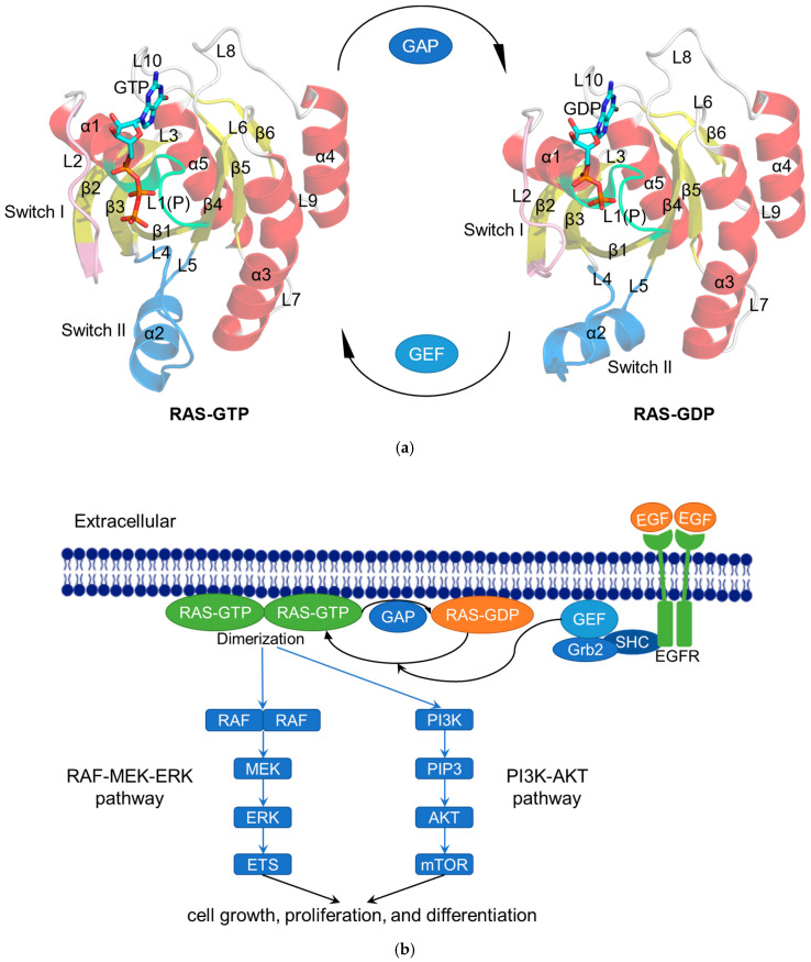
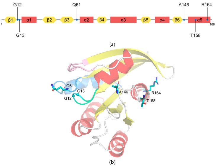
The Rat Sarcoma (RAS) family (NRAS, HRAS, and KRAS) is endowed with GTPase activity to regulate various signaling pathways in ubiquitous animal cells. As proto-oncogenes, RAS mutations can maintain activation, leading to the growth and proliferation of abnormal cells and the development of a variety of human cancers. For the fight against tumors, the discovery of RAS-targeted drugs is of high significance. On the one hand, the structural properties of the RAS protein make it difficult to find inhibitors specifically targeted to it. On the other hand, targeting other molecules in the RAS signaling pathway often leads to severe tissue toxicities due to the lack of disease specificity. However, computer-aided drug design (CADD) can help solve the above problems. As an interdisciplinary approach that combines computational biology with medicinal chemistry, CADD has brought a variety of advances and numerous benefits to drug design, such as the rapid identification of new targets and discovery of new drugs. Based on an overview of RAS features and the history of inhibitor discovery, this review provides insight into the application of mainstream CADD methods to RAS drug design.

Keywords: RAS inhibitor; computer-aided drug design; molecular docking; molecular dynamics simulation; virtual screening.


Conflict of interest statement

The authors declare no conflict of interest.

Figures

Figure 1
Figure 1
The RAS functions as a binary switch in normal state. (a) Cartoon representation of the crystal structure of RAS complexes: KRAS4B–GTP (modified from KRAS4B–GppNHp; PDB ID: 3GFT) and KRAS4B–GDP (PDB ID: 4LPK). The helices (α1–α5), strands (β1–β6), and loops (L1–L10) are shown in red, yellow, and gray, respectively. The P-loop (P or L1), Switch I, and Switch II regions are colored lime, pink, and blue, respectively. GTP/GDP are depicted by stick models [23]. (b) Schematic diagram showing the RAS-related signaling pathways. After activation by epidermal growth factor (EGF), the tyrosine kinase receptor EGFR recruits GEF such as SOS to the cell membrane via Src homology 2 domain containing (SHC) and growth-factor-receptor-bound protein 2 (Grb2) to activate RAS [24]. Subsequently, the activated RAS dimerizes and binds to the downstream RAF protein to regulate the MAPK signaling pathway (RAS–RAF–MEK–ERK pathway). The activated ERK is transported to the nucleus and then phosphorylates a number of transcription factors, such as erythroblastosis virus transcription factor (ETS), to ultimately regulate the cell cycle [25]. In another RAS–PI3K–AKT pathway, the activated RAS recruits PI3K to phosphorylate the substrate PIP2 and generate PIP3, whereupon PIP3 causes the sequential phosphorylation of AKT and mTOR to regulate cell proliferation [26].
Figure 2
Figure 2
Mutations on KRAS. (a) Schematic diagram showing the positions of KRAS mutations; (b) stick representation showing six residue mutations mapped on the cartoon representation of the crystal structure of KRAS.
Figure 3
Figure 3
Surface representation of five potential druggable sites (S1–S3, Subsite 1 and Subsite 2) on KRAS from PMD simulation (PDB ID: 4DSO).
Figure 4
Figure 4
Surface representation of three potential allosteric sites (P1–P3) on RAS from FTMAP.
Figure 5
Figure 5
Surface representation of eight potential binding sites (Cluster 1–Cluster 8) on HRAS from the MSCS method.

Similar articles

Cited by

References

    1. Prior I.A., Hood F.E., Hartley J.L. The Frequency of Ras Mutations in Cancer. Cancer Res. 2020;80:2969–2974. doi: 10.1158/0008-5472.CAN-19-3682. - DOI - PMC - PubMed
    1. Simanshu D.K., Nissley D.V., McCormick F. RAS Proteins and Their Regulators in Human Disease. Cell. 2017;170:17–33. doi: 10.1016/j.cell.2017.06.009. - DOI - PMC - PubMed
    1. Liu S., Alnammi M., Ericksen S.S., Voter A.F., Ananiev G.E., Keck J.L., Hoffmann F.M., Wildman S.A., Gitter A. Practical Model Selection for Prospective Virtual Screening. J. Chem. Inf. Model. 2019;59:282–293. doi: 10.1021/acs.jcim.8b00363. - DOI - PMC - PubMed
    1. Maia E.H.B., Assis L.C., de Oliveira T.A., da Silva A.M., Taranto A.G. Structure-Based Virtual Screening: From Classical to Artificial Intelligence. Front. Chem. 2020;8:343. doi: 10.3389/fchem.2020.00343. - DOI - PMC - PubMed
    1. Yang H., Du Bois D.R., Ziller J.W., Nowick J.S. X-ray crystallographic structure of a teixobactin analogue reveals key interactions of the teixobactin pharmacophore. Chem. Commun. 2017;53:2772–2775. doi: 10.1039/C7CC00783C. - DOI - PMC - PubMed