Review
. 2022 Oct:76:102446.
doi: 10.1016/j.sbi.2022.102446. Epub 2022 Sep 6.

Telomerase structural biology comes of age

Affiliations
Review

Telomerase structural biology comes of age

Yao He et al. Curr Opin Struct Biol. 2022 Oct.

Abstract

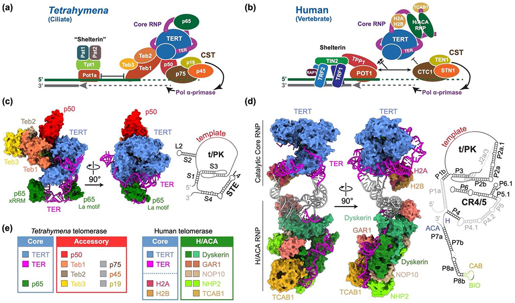
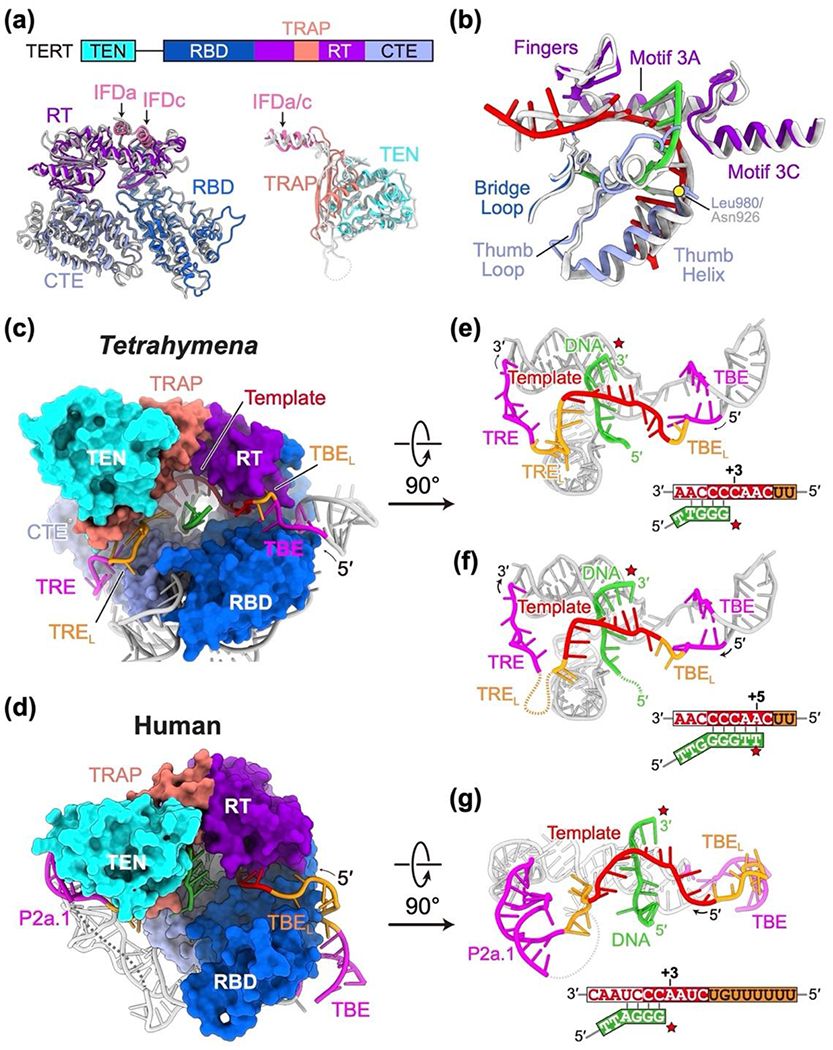
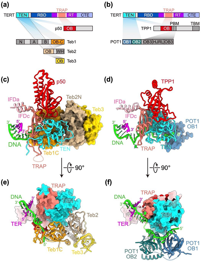
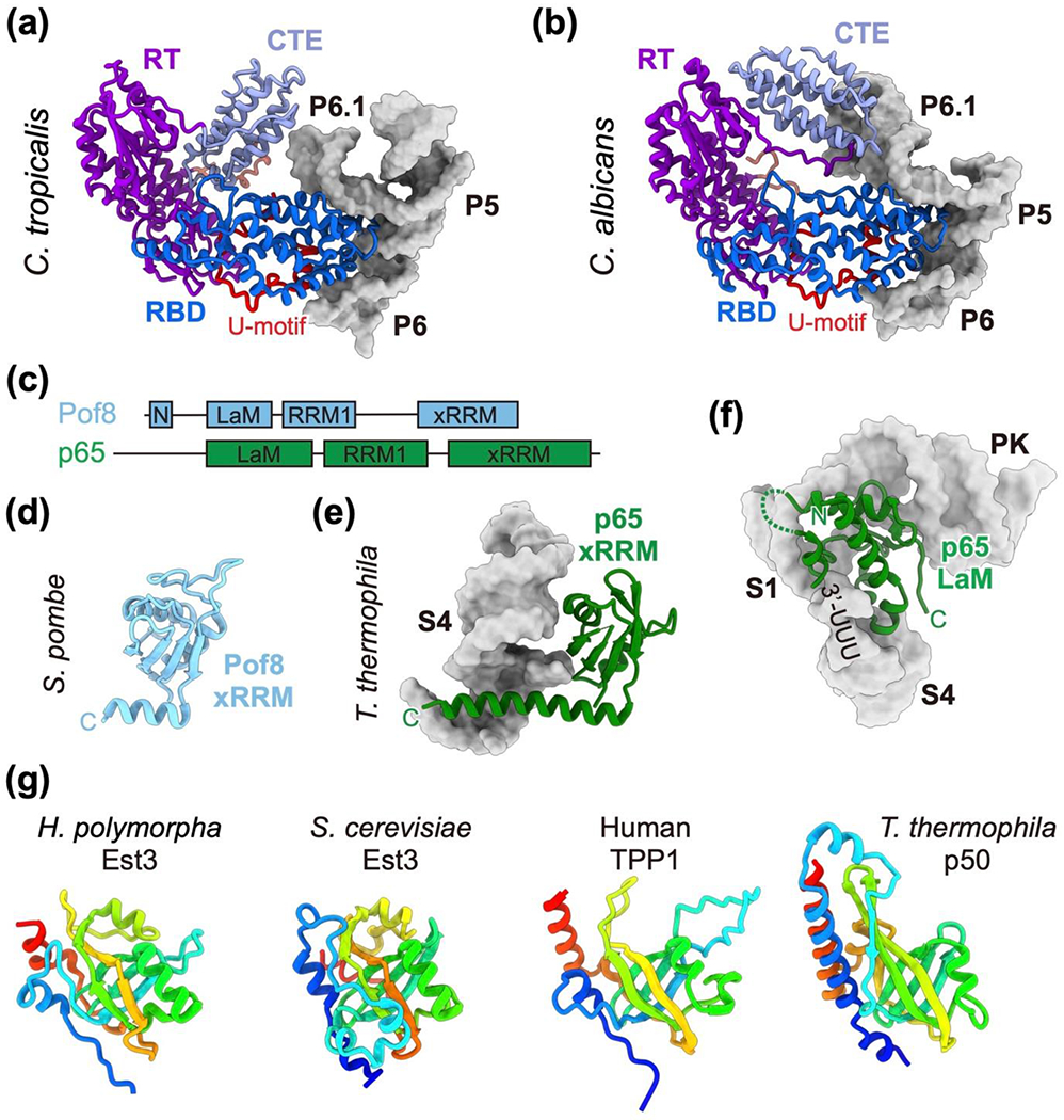
Telomerase is an RNA-protein complex comprising telomerase reverse transcriptase, a non-coding telomerase RNA, and proteins involved in biogenesis, assembly, localization, or recruitment. Telomerase synthesizes the telomeric DNA at the 3'-ends of linear chromosomes. During the past decade, structural studies have defined the architecture of Tetrahymena and human telomerase as well as protein and RNA domain structures, but high-resolution details of interactions remained largely elusive. In the past two years, several sub-4 Å cryo-electron microscopy structures of telomerase were published, including Tetrahymena telomerase at different steps of telomere repeat addition and human telomerase with telomere shelterin proteins that recruit telomerase to telomeres. These and other recent structural studies have expanded our understanding of telomerase assembly, mechanism, recruitment, and mutations leading to disease.


Conflict of interest statement

Conflict of interest statement Nothing declared.

Figures

Figure 1.
Figure 1.. Structures of Tetrahymena and human telomerase holoenzymes.
(a,b) Schematics of Tetrahymena (a) and a human (b) telomerase holoenzymes and their interactions at telomere ends. Tetrahymena telomerase catalytic core RNP is TERT, TER, and p65. p50, TEB (Teb1–Teb2–Teb3, a Replication Protein A (RPA) related complex), and p75–p45–p19 (Tetrahymena Ctc1–Snt1–Ten1, CST) are constitutively associated with the core RNP[79]. Human telomerase catalytic core RNP is TERT, TER, and possibly histone H2A/H2B dimer[8]. The H/ACA RNP has two each Dyskerin, GAR1, NOP10, and NHP2, plus one TCAB1 protein. Human and Tetrahymena CST recruit DNA polymerase α-Primase for synthesis of the C-strand, after G-strand synthesis by telomerase[19]. In human, six proteins collectively called shelterin bind the telomeric DNA[18]; a less well-defined “shelterin” complex of four proteins has been identified in Tetrahymena[80]. (c) 3.3 Å resolution cryo-EM structure of Tetrahymena telomerase (PDB 7LMA), with the dynamic p75–p45–p19 (CST) complex masked out[12], and schematic of TER. For TEB, only the Teb1C, Teb2N, Teb3 heterotrimer is visible; for p50, only the OB-domain is visible; and for p65, only the La motif and xRRM are well defined in the cryo-EM map. (d) Cryo-EM structure of human telomerase (PDB 7TRC for H/ACA RNP and PDB 7TRD for catalytic core RNP) and schematic of TER. Gray colored regions of TER between the two RNPs are modeled based on low resolution cryo-EM densities[8]. (e) Color chart for the telomerase proteins and RNA.
Figure 2.
Figure 2.. Telomerase TERT–TER catalytic core.
(a) Schematic of TERT domains and overlay of human (colored) and Tetrahymena (gray) RBD-RT-CTE TERT ring and TEN–TRAP. (b) Overlay of human (colored) and Tetrahymena (gray) TERT ring showing some motifs that interact with the template and telomeric DNA. (c,d) Surface renderings of telomerase catalytic cavity of Tetrahymena (c) and human (d) with TER and telomeric DNA shown as ribbon-and-sticks. CR4/5 is removed for clarity in (d). Colors for TERT domains are as in (a). (e-g) TER t/PK and telomeric DNA structures in Tetrahymena at the second (e) and fifth (f) steps of telomere repeat addition and in human at the second step of telomere repeat addition (g). Template is red, flexible regions of TER next to template are orange, and fixed regions of TER are magenta. TBE is template boundary element. TBEl is TBE linker. TRE is template recognition element. TREl is TRE linker. In (f) the telomeric DNA is still in the active site, while for (e) and (g) the active site is empty. In the accompanying schematics, red star is active site and numbering refers to template position in the active site for telomere repeat synthesis. The alignment nucleotides in the template are red with white background.
Figure 3.
Figure 3.. Telomerase activation and recruitment complexes.
(a,b) Schematics of domain structures and interactions between (a) Tetrahymena TERT, p50, and TEB (PDB 7LMA) and (b) human TERT, TPP1, and POT1 (PDB 7QXB). Domains that are not visible or defined by the cryo-EM map are shown in gray. (c,d) View of the three-way interactions between Tetrahymena TERT TEN, TRAP, and p50 (c) and human TERT TEN, TRAP, and TPP1 (d), shown in ribbon. TEB (Teb1, Teb2, Teb3) in Tetrahymena (c) and POT1 OB1-OB2 in human (d) are shown as space fill. The short template–telomeric DNA duplex and single-strand exiting telomeric DNA are also shown. (e,f) 90° rotated views of (c,d) showing the path of telomeric DNA in Tetrahymena (e) and human (f).
Figure 4.
Figure 4.. Structural analysis of yeast TERT, Pof8, and Est3.
(a) Structure of C. tropicalis TERT in complex with TER three-way junction (PDB 6ZDP). (b) Structure of C. albicans TERT in complex with TER three-way junction (PDB 6ZDU). Two different conformations of CTE were observed in these two Candida TERT structures. The U-motifs are colored in red. (c) Domain architectures of S. pombe Pof8 and T. thermophila p65. LaM, La motif. (d) Structure of Pof8 xRRM domain (PDB 6TZN and 6U7V). (e) Structure of p65 xRRM domain with TER S4 (PDB 7LMA). (f) Structure of p65 LaM with TER PK, S1, S4 and the 3’-UUU (PDB 7LMA). (g) Structure of H. polymorpha Est3 OB domain (PDB 6Q44) and its comparison with the OB domains of T. thermophila p50 (PDB 7LMA), human TPP1 (PDB 7TRE), and S. cerevisiae Est3 (PDB 2M9V). Structures are rainbow colored from N-terminal (blue) to C-terminal (red).

Similar articles

Cited by

References

    1. Blackburn EH, Collins K: Telomerase: an RNP enzyme synthesizes DNA. Cold Spring Harb Perspect Biol 2011, 3. - PMC - PubMed
    1. Roake CM, Artandi SE: Regulation of human telomerase in homeostasis and disease. Nat Rev Mol Cell Biol 2020, 21:384–397. - PMC - PubMed
    1. Jiang J, Miracco EJ, Hong K, Eckert B, Chan H, Cash DD, Min B, Zhou ZH, Collins K, Feigon J: The architecture of Tetrahymena telomerase holoenzyme. Nature 2013, 496:187–192. - PMC - PubMed
    1. Sauerwald A, Sandin S, Cristofari G, Scheres SH, Lingner J, Rhodes D: Structure of active dimeric human telomerase. Nat Struct Mol Biol 2013, 20:454–460. - PMC - PubMed
    1. Jiang J, Chan H, Cash DD, Miracco EJ, Ogorzalek Loo RR, Upton HE, Cascio D, O’Brien Johnson R, Collins K, Loo JA, et al.: Structure of Tetrahymena telomerase reveals previously unknown subunits, functions, and interactions. Science 2015, 350:aab4070. - PMC - PubMed

Publication types