. 2022:36:103173.
doi: 10.1016/j.nicl.2022.103173. Epub 2022 Aug 29.

Use of magnetic resonance elastography to gauge meningioma intratumoral consistency and histotype

Affiliations

Use of magnetic resonance elastography to gauge meningioma intratumoral consistency and histotype

Yu Shi et al. Neuroimage Clin. 2022.

Abstract

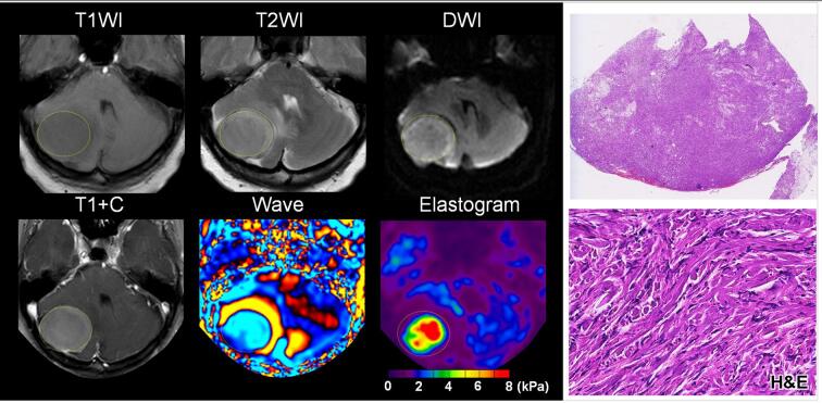
Objective: To determine whether tumor shear stiffness, as measured by magnetic resonance elastography, corresponds with intratumoral consistency and histotype.

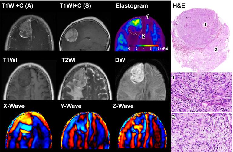
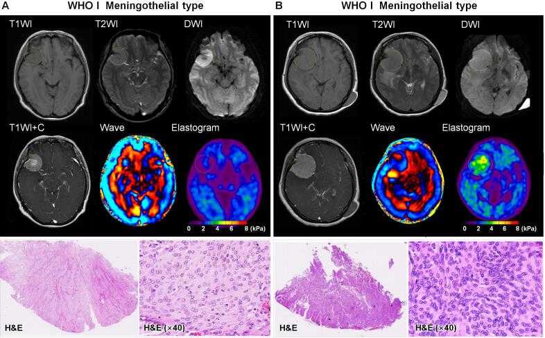
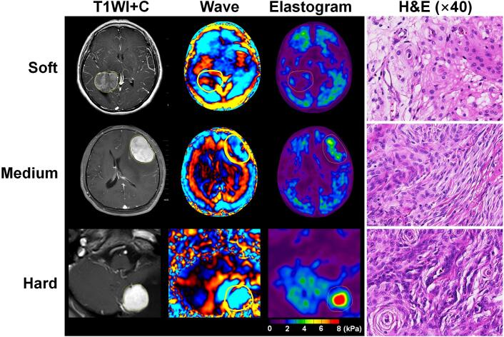
Materials and methods: A total of 88 patients with 89 meningiomas (grade 1, 74 typical [13 fibroblastic, 61 non-fibroblastic]; grade 2, 12 atypical; grade 3, 3 anaplastic) were prospectively studied, each undergoing preoperative MRE in conjunction with T1-, T2- and diffusion-weighted imaging. Contrast-enhanced T1-weighted sequences were also obtained. Tumor consistency was evaluated as heterogeneous or homogenous, and graded on a 5-point scale intraoperatively. MRE-determined shear stiffness was associated with tumor consistency by surgeon's evaluation and whole-slide histologic analyses.

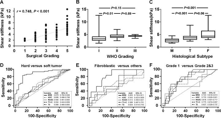
Results: Mean tumor stiffness overall was 3.81+/-1.74 kPa (range, 1.57-12.60 kPa), correlating well with intraoperative scoring (r = 0.748; p = 0.001). MRE performed well as a gauge of tumor consistency (AUC = 0.879, 95 % CI: 0.792-0.938) and heterogeneity (AUC = 0.773, 95 % CI: 0.618-0.813), significantly surpassing conventional MR techniques (DeLong test, all p < 0.001 after Bonferroni adjustment). Shear stiffness was independently correlated with both fibrous content (partial correlation coefficient = 0.752; p < 0.001) and tumor cellularity (partial correlation coefficient = 0.547; p < 0.001). MRE outperformed other imaging techniques in distinguishing fibroblastic meningiomas from other histotypes (AUC = 0.835 vs 0.513 ∼ 0.634; all p < 0.05), but showed limited ability to differentiate atypical or anaplastic meningiomas from typical meningiomas (AUC = 0.723 vs 0.616 ∼ 0.775; all p > 0.05). Small (<2.5 cm, n = 6) and intraventricular (n = 2) tumors displayed inconsistencies between MRE and surgeon's evaluation.

Conclusions: The results of this prospective study provide substantial evidence that preoperative evaluation of meningiomas with MRE can reliably characterize tumor stiffness and spatial heterogeneity to aid neurosurgical planning.

Keywords: Elastography; Magnetic resonance imaging; Mechanical properties; Meningiomas; Stiffness.


Conflict of interest statement

Declaration of Competing Interest The Mayo Clinic and RLE have intellectual property rights and a financial interest related to MRE technology.

Figures

Fig. 1
Fig. 1
(A) Spearman’s correlation test, indicating strong correlation between shear stiffness level and grading by surgeon (r = 0.748; p < 0.001); (B) Shear stiffness of meningiomas stratified by World Health Organization (WHO) grade, pairwise comparisons (Mann-Whitney U test) showing significant difference between grades 1 and 2 (p = 0.01), but not between grades 1 and 3 (p = 0.15) or grades 2 and 3 (p = 0.89); (C) Shear stiffness of three main WHO grade 1 meningioma histotypes (T, transitional; M, meningothelial; F, fibroblastic), pairwise comparisons indicating significant difference in mean stiffness for T vs M (p < 0.001) and for M vs F (p < 0.001), but not for T vs F (p = 0.06); (D-F) Composite graph of receiver operating characteristic (ROC) curves comparing performance of shear stiffness with other conventional imaging techniques in identifying firm, fibroblastic, and grade 2–3 meningiomas, respectively. Traditional imaging measures included tumor apparent diffusion coefficient (ADC), relative ADC (mass ADC/brain ADC), signal intensity on T2-weighted imaging and contrast-enhanced T1-weighted imaging.
Fig. 2
Fig. 2
Representative images of World Health Organization grade 1 transitional meningioma at right frontal lobe, heterogenous in consistency (area 1: anterior portion; area 2: posterior portion). Conventional contrast-enhanced MR shows two distinctive areas of similar signal intensity on T1-weighted, T2-weighted, diffusion-weighted, and contrast-enhanced images, whereas MRE indicates tumor heterogeneity, firmer anteriorly (area 1: 3.2 ± 0.24 kPa) and softer posteriorly (area 2: 1.6 ± 0.34 kPa). Hematoxylin-eosin staining (original magnification, 40x) confirms greater tumor cellularity and fibrous content in area 1.
Fig. 3
Fig. 3
(A) 48-year-old woman with World Health Organization (WHO) grade 1 meningothelial meningioma of right frontotemporal lobe. Pre-contrast T1- and T2-weighted MR (magnetic resonance) images show a mostly homogenous, isointense, round tumor with clear border. Diffusion-weighted images reveal hyperintensity. In T1-weighted MR views, there is ready enhancement, particularly at central core. Shear stiffness by MR elastography was 1.85 ± 0.32 kPa. Intraoperatively, the tumor was easily suctioned and consistent throughout. In hematoxylin & eosin (HE)-stained sections, the neoplastic cells closely resemble arachnoid cap cells, displaying round or oval, centrally placed nuclei and abundant cytoplasm. Cellularity is low level (986 cells, 40x magnification); (B) 53-year-old woman with WHO grade 1 meningothelial meningioma, again in right frontotemporal lobe. Similar signal intensity is evident on pre-contrast T1- and T2-weighted images. Diffusion-weighted imaging shows lower signal intensity than the tumor above and more homogeneous enhancement. MR elastography indicates a stiffer tumor (3.87 ± 0.72 kPa). Intraoperatively, the tumor required some degree of mechanical debulking, cauterization and full decompression. In HE-stained sections, neoplastic cells resembled those above, with approximately twice the density (2120 cells, 40x magnification).
Fig. 4
Fig. 4
Images of three World Health Organization (WHO) grade 1 transitional meningiomas that differ in consistency. Transitional meningiomas contain a mix of meningothelial and fibrous elements, here demonstrating meningothelial cells in fascicles of variable length, with fibroblastic appearance and syncytial pattern. The soft tumor (top row, 1.7 ± 0.3 kPa) of right lateral ventricle had a cell count of 713 (40x) and 20 % fibrous content; the tumor of medium consistency (middle row, 2.8 ± 0.45 kPa) in left frontal lobe appears more densely cellular, with 1543 cells (40x) and 37 % fibrous tissue; and the firm tumor (bottom row, 6.8 ± 0.74 kPa) of left cerebellum bears the densest meningothelial population, with 1843 cells (40x) and 44 % fibrous content.
Fig. 5
Fig. 5
Representative images from a 52-year-old man with World Health Organization (WHO) grade 1 fibroblastic meningioma of right cerebellum. Pre-contrast MR studies showed slightly low signal intensity on T1-weighted imaging, isointense signal on T2-weighted imaging, slightly hyperintense signal intensity on diffusion-weighted imaging, and moderate homogenous enhancement. By MR (magnetic resonance) elastography, the tumor was firm and homogenous (7.1 ± 0.7 kPa). Intraoperatively, tumor composition was consistent throughout, requiring cautery for complete removal. In HE-stained sections, this firm fibrous meningioma was composed of spindle cells with narrow, rod-shaped nuclei and indistinct cell boundaries, with an abundance of collagen or reticulin in the background.
Fig. 6
Fig. 6
Representative images from a 60-year-old man with World Health Organization (WHO) grade 3 meningioma of right frontotemporal lobe. There was heterogenous tumor signal intensity on all images (4.24 ± 1.2 kPa), mostly isointense on T1- and T2-weighted imaging and hyperintense on diffusion-weighted imaging. Ready heterogenous enhancement was observed on axial contrast-enhanced T1-weighted imaging, with signs of necrosis. Photomicrographs of the whole-slide digital image and slides digitized at 200x magnification showed a dense array of spindle-shaped cell in fascicles, along with necrosis (dashed black rectangle). Some tumor cells varied in size, showing moderate atypia. Mitotic activity in cell-dense areas was ∼ 5 per 10 high-power fields (HPFs). The cytoplasm stained positive for epithelial membrane antigen (EMA), partial nuclear positivity for progesterone receptor (PR) was observed, and Ki-67 was positive over ∼ 60 % of a hot spot.

References

    1. Alyamany M., Alshardan M.M., Jamea A.A., ElBakry N., Soualmi L., Orz Y. Meningioma Consistency: Correlation Between Magnetic Resonance Imaging Characteristics, Operative Findings, and Histopathological Features. Asian J. Neurosurg. 2018;13:324–328. - PMC - PubMed
    1. Backer-Grondahl T., Moen B.H., Torp S.H. The histopathological spectrum of human meningiomas. Int. J. Clin. Exp. Pathol. 2012;5:231–242. - PMC - PubMed
    1. Cepeda S., Arrese I., Garcia-Garcia S., Velasco-Casares M., Escudero-Caro T., Zamora T., Sarabia R. Meningioma Consistency Can Be Defined by Combining the Radiomic Features of Magnetic Resonance Imaging and Ultrasound Elastography. A Pilot Study Using Machine Learning Classifiers. World Neurosurg. 2021;146:e1147–e1159. - PubMed
    1. Chakraborty A., Bamber J.C., Dorward N.L. Slip elastography: a novel method for visualising and characterizing adherence between two surfaces in contact. Ultrasonics. 2012;52(3):364–376. - PubMed
    1. Chen T.C., Zee C.S., Miller C.A., Weiss M.H., Tang G., Chin L., Levy M.L., Apuzzo M.L. Magnetic resonance imaging and pathological correlates of meningiomas. Neurosurgery. 1992;31:1015–1021. discussion 1021–1012. - PubMed

MeSH terms