Comment
. 2022 Aug 16:11:22.
doi: 10.12703/r-01-0000015. eCollection 2022.

An alliance between lipid transfer proteins and scramblases for membrane expansion

Affiliations
Comment

An alliance between lipid transfer proteins and scramblases for membrane expansion

Joost C M Holthuis et al. Fac Rev. .

Abstract

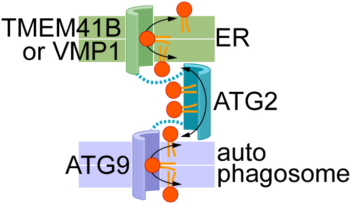
Membrane growth requires lipid supply, which is usually accomplished by lipid synthesis or vesicular trafficking. In the case of autophagosomes, these principles do not apply. Ghanbarpour et al. postulate that autophagosome expansion relies on non-vesicular lipid delivery from the ER, whereby the activity of a lipid transfer protein (LTP) is directly coupled to scramblase activities in the donor and acceptor bilayers1. This new concept opens the possibility that lipid traffic is controlled by scramblases that provide not only specific docking sites for LTPs, thereby directing lipid flow, but also support their activity by overcoming barriers for lipid extraction and deposition.

Keywords: Scramblase; autophagosome; lipid; lipid transfer protein; membrane.


Conflict of interest statement

The authors declare that they have no competing interests.

Figures

Figure 1
Figure 1. Proposed cooperation between scramblases in the ER (TMEM41B, VMP1) and nascent autophagosomal membrane (ATG9), and the bridging lipid transport protein ATG2
The membrane bilayers are shown as coloured slabs (a white line separates the two halves of each bilayer). Phospholipids are shown generically with a red headgroup and orange acyl chains. Docking of ATG2 to the scramblases is indicated by the dotted lines. The transport proteins are shown according to the credit card model, with a polar groove in the case of scramblases to accommodate lipid headgroups, and a hydrophobic groove in ATG2 to accommodate lipid tails. Bidirectional flow of lipids is shown by double-headed arrows so that lipids in both leaflets of both membranes are equilibrated by the scramblases and inter-bilayer exchange across the cytoplasm. Lipid transport may be effectively one-directional, with lipids being synthesized in the ER (‘source’) and consumed through expansion of the autophagosomal membrane (‘sink’) (see ‘Open Questions’ section ‘What drives lipids to move towards the acceptor membrane, i.e., the newly forming autophagosome membrane’ for details).

Comment on

References

    1. Ghanbarpour A, Valverde DP, Melia TJ, Reinisch KM. 2021. A model for a partnership of lipid transfer proteins and scramblases in membrane expansion and organelle biogenesis Proc Natl Acad Sci U S A 118:e2101562118. 10.1073/pnas.2101562118 - DOI - PMC - PubMed
    2. Faculty Opinions Recommendation

    1. Osawa T, Kotani T, Kawaoka T, Hirata E, Suzuki K, Nakatogawa H, Ohsumi Y, Noda NN. 2019. Atg2 mediates direct lipid transfer between membranes for autophagosome formation Nat Struct Mol Biol 26:281–288. 10.1038/s41594-019-0203-4 - DOI - PubMed
    2. Faculty Opinions Recommendation

    1. Valverde DP, Yu S, Boggavarapu V, Kumar N, Lees JA, Walz T, Reinisch KM, Melia TJ. 2019. ATG2 transports lipids to promote autophagosome biogenesis J Cell Biol 218:1787–1798. 10.1083/jcb.201811139 - DOI - PMC - PubMed
    2. Faculty Opinions Recommendation

    1. Maeda S, Otomo C, Otomo T. 2019. The autophagic membrane tether ATG2A transfers lipids between membranes eLife 8:e45777. 10.7554/eLife.45777 - DOI - PMC - PubMed
    2. Faculty Opinions Recommendation

    1. Wong LH, Gatta AT, Levine TP. 2019. Lipid transfer proteins: the lipid commute via shuttles, bridges and tubes Nat Rev Mol Cell Biol 20:85–101. 10.1038/s41580-018-0071-5 - DOI - PubMed

LinkOut - more resources