Review
. 2022 Aug 19:10:933474.
doi: 10.3389/fcell.2022.933474. eCollection 2022.

Mechanical regulation of signal transduction in angiogenesis

Affiliations
Review

Mechanical regulation of signal transduction in angiogenesis

Jennifer Flournoy et al. Front Cell Dev Biol. .

Abstract

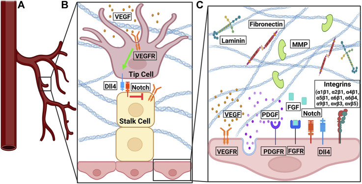
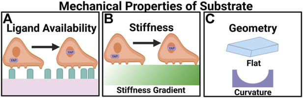
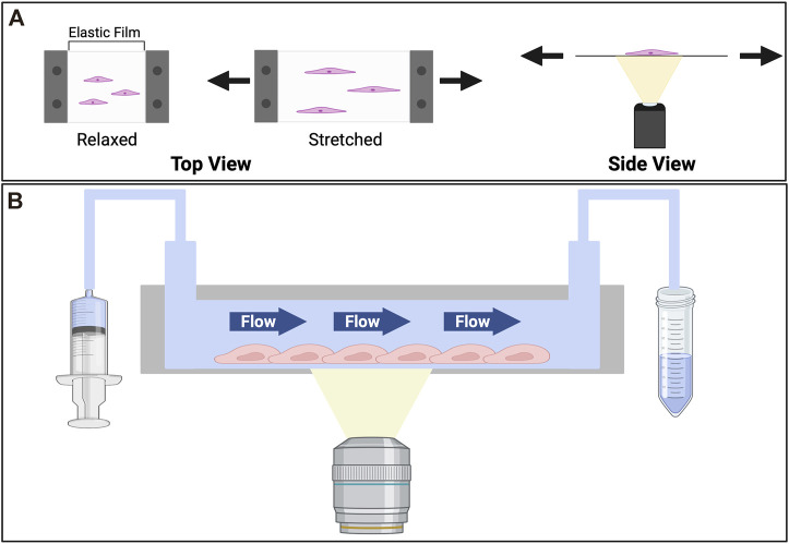
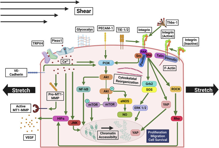
Biophysical and biochemical cues work in concert to regulate angiogenesis. These cues guide angiogenesis during development and wound healing. Abnormal cues contribute to pathological angiogenesis during tumor progression. In this review, we summarize the known signaling pathways involved in mechanotransduction important to angiogenesis. We discuss how variation in the mechanical microenvironment, in terms of stiffness, ligand availability, and topography, can modulate the angiogenesis process. We also present an integrated view on how mechanical perturbations, such as stretching and fluid shearing, alter angiogenesis-related signal transduction acutely, leading to downstream gene expression. Tissue engineering-based approaches to study angiogenesis are reviewed too. Future directions to aid the efforts in unveiling the comprehensive picture of angiogenesis are proposed.

Keywords: ECM; angiogenesis; mechanotransduction; shear stress; stiffness; stretch.


Conflict of interest statement

The authors declare that the research was conducted in the absence of any commercial or financial relationships that could be construed as a potential conflict of interest.

Figures

FIGURE 1
FIGURE 1
The signaling pathways involved in angiogenesis. (A) At the front of a sprouting vessel, (B) VEGF binds to VEGFR on tip cells. VEGFR activation induced the expression of Dll4. Dll4 on the tip cells binds to Notch on stalk cells. Notch signaling in stalk cells downregulates the expression of VEGFR2/3 in stalk cells. (C) VEGF-VEGFR and FGF-FGFR signaling promote pro-angiogenic signals, cell proliferation, and cell survival. PDGF-PDGFR binding leads to vessel sprouting and stabilization. Notch interacts with Dll4 in a feedback loop to regulate cell migration and proliferation. Fibronectin and laminin bind to integrins to promote endothelial cell proliferation, survival, migration, and tube formation. Various integrin isoforms promote angiogenesis through different overlapping pathways. MMPs degrade denatured collagen in the basement membrane.
FIGURE 2
FIGURE 2
Angiogenesis is regulated by the mechanical properties of the microenvironment. (A) Ligand availability regulates the migratory behavior of cells through haptotaxis. In high ligand densities YAP translocates to the nucleus. (B) Greater stiffness upregulates the expression of pro-angiogenic factors. On stiff substrates YAP undergoes nuclear localization. (C) Appropriate curvatures of the substrates can promote angiogenesis.
FIGURE 3
FIGURE 3
Devices for in vitro studies of effects imposed by the tensile stress (stretch) and shear stress (flow). (A) Cell stretcher. To observe the effects of stretch cells are cultured on an elastic substrate which is mounted on a cell stretching device. The stretching device that reversibly deforms the elastic film. The stretcher itself is commonly mounted on an inverted microscope, where an objective underneath the elastic slide facilitates the imaging of dynamic cell behaviors upon stretching. To stretch the cells a prescribed strain is applied to the elastic substrate. (B) Microfluidic device. Consisting of a cell culture chamber with an inlet connecting to a syringe pump, and an outlet to collect the flow-through, the device can be used to study cellular responses to shear stress. Cells are seeded in the chamber. Medium is flown through at a prescribed rate. To collect images of cells over time, the device usually is mounted on an inverted microscope, where an objective is placed beneath the cell culture chamber.
FIGURE 4
FIGURE 4
Mechanosensitive pathways involved in signaling triggered by stretching and/or shear stress. Ca2+ influx through Piezo1 and TRPV4 upregulates MT1-MMP, PI3K, JNK, promoting cell growth and proliferation, and HIFα, upregulating VEGF expression. PI3K is also upregulated by PECAM-1, integrin, and VE-Cadherin, which are activated by mechanical stress, and in turn, activate Akt. Akt’s downstream pathways activate NF-κB and mTOR, which facilitate cell growth and proliferation, as well as eNOS and NO, which enhance cell survival. Activated integrin, to which a macromolecular complex of FAK, Shc, Src, and Paxillin is bound, interacts with TIE-1/2. Rho and ROCK are activated by FAK and/or Src, upregulating in HIFα. Shc upregulates Grb2, SOS, and FRK 1/2, promoting cell proliferation and growth. Under shear stress and stretch YAP is translocated to the nucleus.

References

    1. Abhinand C. S., Raju R., Soumya S. J., Arya P. S., Sudhakaran P. R. (2016). VEGF-A/VEGFR2 signaling network in endothelial cells relevant to angiogenesis. J. Cell Commun. Signal. 10, 347–354. 10.1007/s12079-016-0352-8 - DOI - PMC - PubMed
    1. Acerbi I., Cassereau L., Dean I., Shi Q., Au A., Park C., et al. (2015). Human breast cancer invasion and aggression correlates with ECM stiffening and immune cell infiltration. Integr. Biol. (United Kingdom) 7, 1120–1134. 10.1039/c5ib00040h - DOI - PMC - PubMed
    1. Ades E. W., Candal F. J., Swerlick R. A., George V. G., Summers S., Bosse D. C., et al. (1992). HMEC-1: Establishment of an immortalized human microvascular endothelial cell line. J. Invest. Dermatol. 99, 683–690. 10.1111/1523-1747.ep12613748 - DOI - PubMed
    1. Ahn G. O., Brown J. M. (2009). Influence of bone marrow-derived hematopoietic cells on the tumor response to radiotherapy: Experimental models and clinical perspectives. Cell Cycle 8, 970–976. 10.4161/cc.8.7.8075 - DOI - PMC - PubMed
    1. Akbari E., Spychalski G. B., Rangharajan K. K., Prakash S., Song J. W. (2019). Competing fluid forces control endothelial sprouting in a 3-D microfluidic vessel bifurcation model. Micromachines 10, 451. 10.3390/mi10070451 - DOI - PMC - PubMed

LinkOut - more resources