. 2022 Aug 23:16:974692.
doi: 10.3389/fnbeh.2022.974692. eCollection 2022.

Early life stress exacerbates behavioural and neuronal alterations in adolescent male mice lacking methyl-CpG binding protein 2 (Mecp2)

Affiliations

Early life stress exacerbates behavioural and neuronal alterations in adolescent male mice lacking methyl-CpG binding protein 2 (Mecp2)

Jose Vicente Torres-Pérez et al. Front Behav Neurosci. .

Abstract

The methyl-CpG binding protein 2 gene (MECP2) encodes an epigenetic transcriptional regulator implicated in neuronal plasticity. Loss-of-function mutations in this gene are the primary cause of Rett syndrome and, to a lesser degree, of other neurodevelopmental disorders. Recently, we demonstrated that both Mecp2 haploinsuficiency and mild early life stress decrease anxiety-like behaviours and neuronal activation in brain areas controlling these responses in adolescent female mice. Here, we extend this work to males by using Mecp2-null and wild type adolescent mice subjected to maternal separation and their non-stressed controls. We assessed their behavioural responses in a battery of anxiety-provoking tests. Upon exposure to an elevated plus maze in aversive conditions, we evaluated changes in c-FOS expression in stress- and anxiety-related brain regions. In addition, we assessed the impact of maternal separation in neuronal maturation using doublecortin and reelin as surrogate markers. Mutant males showed reduced motor abilities, increased activation of the olfactory bulbs, probably due to breathing abnormalities, and decreased activation of the paraventricular thalamic nucleus, when compared to wild type mice. In addition, maternal separation increased the number of immature doublecortin-like neurons found in Mecp2-null animals. Moreover, this work shows for the first time that reelin is decreased in the mutant animals at the olfactory tubercle, piriform cortex and hippocampal dentate gyrus, an effect also associated to maternal separation. Taken together, our results suggest that maternal separation exacerbates some phenotypical alterations associated with lack of MeCP2 in adolescent males.

Keywords: Rett sydrome; c-FOS; doublecortin; maternal separation; reelin.


Conflict of interest statement

The authors declare that the research was conducted in the absence of any commercial or financial relationships that could be construed as a potential conflict of interest.

Figures

FIGURE 1
FIGURE 1
Diagram of the experimental design. In the litters assigned to maternal separation (MS), pups were separated from the dam for 3 h per day between postnatal days (PND) 3 and 21. After MS, pups were returned to their home cage. Behavioural testing started at 5 weeks of age with at least 24 h gap between tests consisting on: elevated plus maze (EPM), open field (OF), forced swim test (FST). The day after FST, animals were subjected to a new EPM but in bright light (more aversive) and 1 h after this test animals were transcardially perfused to obtain brain samples for histological experiments.
FIGURE 2
FIGURE 2
Adolescent Mecp2-null males show motor deficits and reduced anxiety- and depressive-like behavioural responses. Males from both genotypes, WT and Mecp2-null, from either MS or naïve conditions were weighted and tested sequentially for EPM, OF and FST (WT-naïve, n = 15; WT-MS, n = 15, Mecp2-null-naïve, n = 9; Mecp2-null-MS, n = 15). (A) Body weight of the animals. In the EPM, (B) percentage of time in the open arms of the maze, and (C) distance travelled. In the OF, (D) percentage of time spent in the centre of the arena, and (E) total distance travelled. Time spent immobile (F). In the FST, (G) mutant mice spend significantly less time immobile than their WT counterparts. One week after previous behavioural assays, WT and Mecp2-null male mice, from either MS or naïve conditions were re-exposed to EPM under bright light conditions. (H) Time in the open arms of the maze and (I) Distance travelled. Graph show individual values and mean ± standard error mean (SEM) for each group. *p < 0.05, ***p < 0.001 by genotype; +p < 0.05 by group. EPM, elevated plus maze; FST, forced swimming test; MS, maternal separation; OF, open field; WT, wild-type.
FIGURE 3
FIGURE 3
Exposure to the EPM in bright conditions leads to differential c-FOS activation at multiple brain areas that is genotype- and group-dependent. One hour after the re-exposure to the EPM behavioural test under bright conditions, a batch of animals from each genotype x treatment were culled to assess c-FOS immunoreactivity (c-FOS-ir; n = 3-6, depending on genotype/treatment/area). Representative photomicrographs from c-FOS immunostaining at the Pir from (A) WT-naïve, (A’) WT-MS, (A”) Mecp2-null-naïve, (A”’) Mecp2-null-MS. Scale bar represents 100 μm. Principal component analysis of c-FOS expression profiles at the different areas studied are coloured according to (B) genotype, (C) group or (D) both; (E) arrows displaying the weight of the variables (brain areas) for both first and second principal components. Graphs show individual values and mean for each group. DG, dentate gyrus of the hippocampus; OB, olfactory bulb; PA, paraventricular hypothalamic nucleus; PV, paraventricular thalamic nucleus; Pir, piriform cortex; LSV, ventral part of the lateral septum; EPM, elevated plus maize; MS, maternal separation; WT, wild-type.
FIGURE 4
FIGURE 4
c-FOS-ir at multiple brain areas is dependent on Mecp2 dosage and MS. Number of c-FOS positive cells per section at the (A) OB, (B) layer 2 of the Pir, (C) LSV, (D) DG of the hippocampus, (E) PV, and (F) Pa. All graphics describe individual values and mean ± SEM. *p < 0.05 by genotype; +p < 0.05 by group. DG, dentate gyrus of the hippocampus; OB, olfactory bulb; PA, paraventricular hypothalamic nucleus; PV, paraventricular thalamic nucleus; Pir, piriform cortex; LSV, ventral part of the lateral septum.
FIGURE 5
FIGURE 5
Lack of Mecp2 increases the number of DCX positive cells in a region-specific manner and MS further exacerbates this effect. (A) Representative photomicrographs from DCX immunostaining (red) at the Pir from WT-naïve (A), WT-naïve, WT-MS (A’), Mecp2-null-naïve (A”) and Mecp2-null-MS at 10× (left panel) (scale bar 100 μm) and a higher magnification at 20× (right panel) (scale bar 25 μm) Arrow points to a complex and arrowhead to a DCX-ir tangled representative DCX-ir neurons. In the OB: density of DCX-ir cells at the PGL (B) and OD for DCX at the Gr (C). (D) DCX-ir cells in OT and (E). total density of DCX-ir in the Pir, and in the tangled (E’) and complex DCX-ir cells only (E”). Number of DCX positive cells at (F) dSt and (G) vSt. Number of DCX-ir cells in the DG when comparing animals from the MS groups (H). All graphs show individual values and mean ± SEM. *p < 0.05 by genotype; +p < 0.05, ++p < 0.01 by group. DCX, doublecortin; DG, dentate gyrus of the hippocampus; dSt, dorsal striatum; Gr, granular layer of the olfactory bulb; OB, olfactory bulb; OT, olfactory tubercle; PGL, periglomerular layer of the olfactory bulb; Pir, piriform cortex; vSt, ventral striatum.
FIGURE 6
FIGURE 6
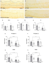
Both MS and the lack of Mecp2 reduce the expression of reelin. (A) Representative photomicrographs from reelin immunostaining (dark-brown, revealed with DAB) at the Pir from WT-naïve (A), WT-MS (A’), Mecp2-null-naïve (A”) and Mecp2-null-MS (A”’); scale bar represents 100 μm. Note that while the positive signal in the Pir layer 1 is restricted to a population of cellular bodies, the distribution in Pir layer 2 is diffuse. Reelin-ir in the OB (B) and the PGL (C). Number of reelin immunopositive cells per section at the OT (D) and layer 1 of the Pir (E) and reelin-ir in layer 2 of the Pir (F). Reelin-ir cells in the (G) GrDG and (H) PoDG. In all cases, graphs represent individual values and mean ± SEM. *p < 0.05, **p < 0.01 by genotype; +p < 0.05, ++p < 0.01 by group. GrDG, granular cell layer of the dentate gyrus of the hippocampus, OT, olfactory tubercle; PGL, periglomerular layer of the olfactory bulb; Pir, piriform cortex; PoDG, polymorph cell layer of the dentate gyrus of the hippocampus.

Similar articles

Cited by

References

    1. Abellán-Álvaro M., Stork O., Agustín-Pavón C., Santos M. (2021). MeCP2 haplodeficiency and early-life stress interaction on anxiety-like behavior in adolescent female mice. J. Neurodev. Disord. 13:59. 10.1186/s11689-021-09409-7 - DOI - PMC - PubMed
    1. Amir R. E., Van den Veyver I. B., Wan M., Tran C. Q., Francke U., Zoghbi H. Y. (1999). Rett syndrome is caused by mutations in X-linked MECP2, encoding methyl-CpG-binding protein 2. Nat. Genet. 23 185–188. 10.1038/13810 - DOI - PubMed
    1. Armario A. (2021). The forced swim test: Historical, conceptual and methodological considerations and its relationship with individual behavioral traits. Neurosci. Biobehav. Rev. 128 74–86. 10.1016/j.neubiorev.2021.06.014 - DOI - PubMed
    1. Barnes K. V., Coughlin F. R., O’Leary H. M., Bruck N., Bazin G. A., Beinecke E. B., et al. (2015). Anxiety-like behavior in Rett syndrome: Characteristics and assessment by anxiety scales. J. Neurodev. Disord. 7:30. 10.1186/s11689-015-9127-4 - DOI - PMC - PubMed
    1. Barney C. C., Feyma T., Beisang A., Symons F. J. (2015). Pain experience and expression in Rett syndrome: Subjective and objective measurement approaches. J. Dev. Phys. Disabil. 27 417–429. 10.1007/s10882-015-9427-3 - DOI - PMC - PubMed

LinkOut - more resources