. 2022 Aug 23:16:964644.
doi: 10.3389/fnbeh.2022.964644. eCollection 2022.

Structural and functional organization of the midline and intralaminar nuclei of the thalamus

Affiliations

Structural and functional organization of the midline and intralaminar nuclei of the thalamus

Robert P Vertes et al. Front Behav Neurosci. .

Abstract

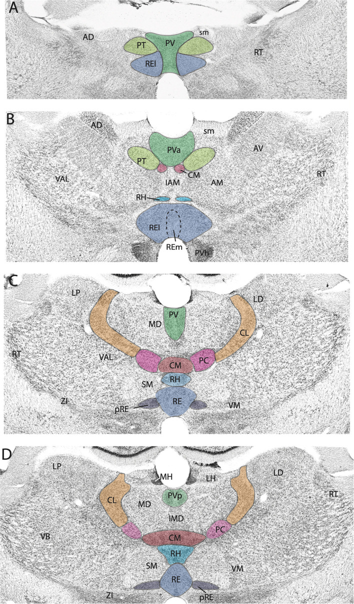
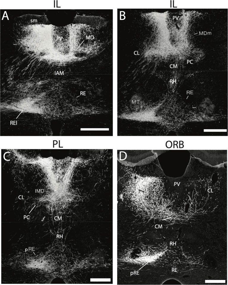
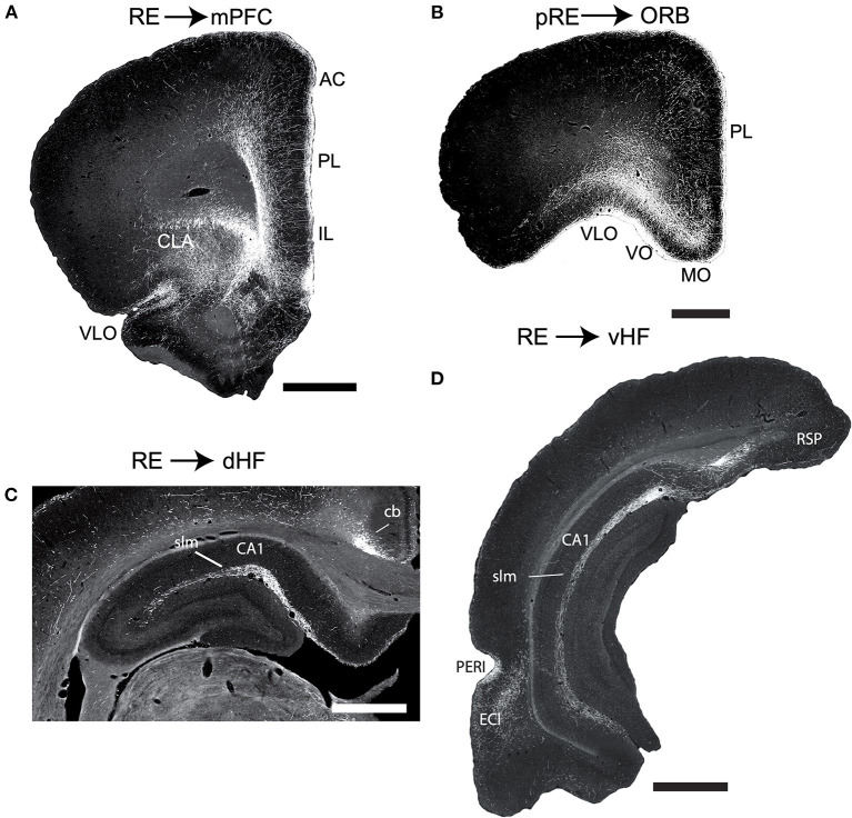
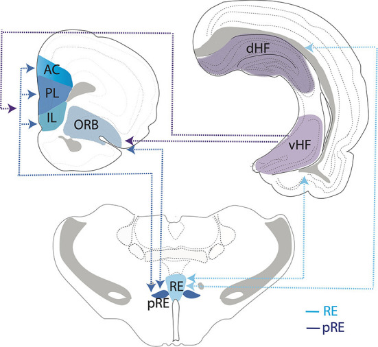
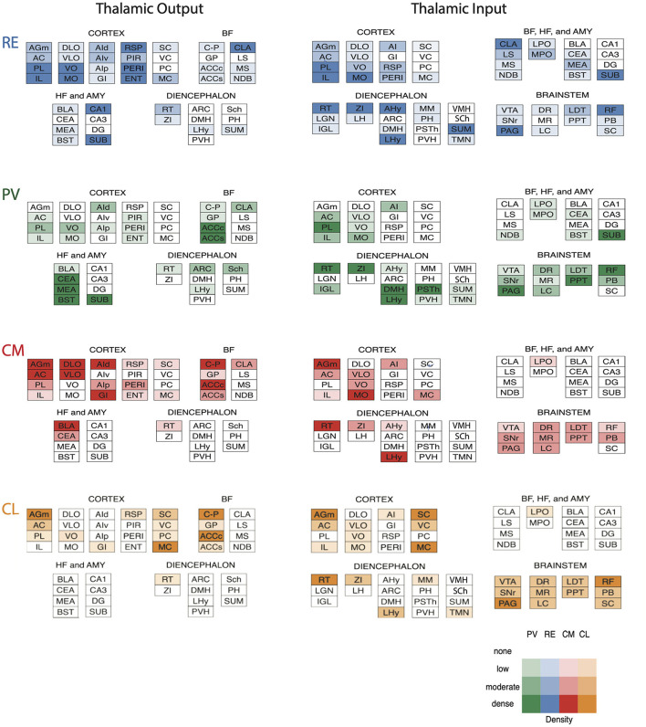
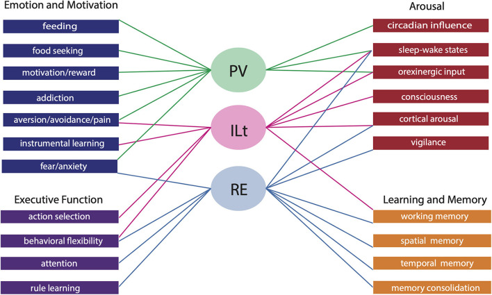
The midline and intralaminar nuclei of the thalamus form a major part of the "limbic thalamus;" that is, thalamic structures anatomically and functionally linked with the limbic forebrain. The midline nuclei consist of the paraventricular (PV) and paratenial nuclei, dorsally and the rhomboid and nucleus reuniens (RE), ventrally. The rostral intralaminar nuclei (ILt) consist of the central medial (CM), paracentral (PC) and central lateral (CL) nuclei. We presently concentrate on RE, PV, CM and CL nuclei of the thalamus. The nucleus reuniens receives a diverse array of input from limbic-related sites, and predominantly projects to the hippocampus and to "limbic" cortices. The RE participates in various cognitive functions including spatial working memory, executive functions (attention, behavioral flexibility) and affect/fear behavior. The PV receives significant limbic-related afferents, particularly the hypothalamus, and mainly distributes to "affective" structures of the forebrain including the bed nucleus of stria terminalis, nucleus accumbens and the amygdala. Accordingly, PV serves a critical role in "motivated behaviors" such as arousal, feeding/consummatory behavior and drug addiction. The rostral ILt receives both limbic and sensorimotor-related input and distributes widely over limbic and motor regions of the frontal cortex-and throughout the dorsal striatum. The intralaminar thalamus is critical for maintaining consciousness and directly participates in various sensorimotor functions (visuospatial or reaction time tasks) and cognitive tasks involving striatal-cortical interactions. As discussed herein, while each of the midline and intralaminar nuclei are anatomically and functionally distinct, they collectively serve a vital role in several affective, cognitive and executive behaviors - as major components of a brainstem-diencephalic-thalamocortical circuitry.

Keywords: affect; arousal; cognition; hippocampus; limbic thalamus; medial prefrontal cortex; striatum.


Conflict of interest statement

The authors declare that the research was conducted in the absence of any commercial or financial relationships that could be construed as a potential conflict of interest.

Figures

Figure 1
Figure 1
(A–D) Nissl-stained micrographs of transverse sections through the diencephalon of the rat depicting nuclei of the thalamus at four anterior to mid-levels of the thalamus. Colored-coded sections show the locations of the midline and rostral intralaminar nuclei of the thalamus. The midline nuclei consist of the paraventricular (PV) and paratenial (PT) of the dorsal midline thalamus and the rhomboid (RH) and reuniens (RE) nuclei of the ventral midline thalamus. The rostral intralaminar nuclei consists of the central medial (CM), paracentral (PC), and central lateral (CL) nuclei. AD, anterodorsal nucleus of thalamus; AM, anteromedial nucleus of thalamus; AV, anteroventral nucleus of thalamus; IAM, interanteromedial nucleus of thalamus; IMD, interomediodorsal nucleus of thalamus; LD, laterodorsal nucleus of thalamus; LH, lateral habenula; LP, lateral posterior nucleus of thalamus; MD, mediodorsal nucleus of thalamus; MH, medial habenula; pRE, peri-reuniens; PVa, anterior PV of thalamus; PVh, paraventricular nucleus of hypothalamus; PVp, posterior PV of thalamus; RE, nucleus reuniens, REl, REm, lateral, medial division of RE; RT, reticular nucleus of thalamus; sm, stria medullaris; SM, submedial nucleus of thalamus; VAL, ventral anterior nucleus of thalamus; VB, ventrobasal nucleus of thalamus; VM, ventromedial nucleus of thalamus; ZI, zona incerta.
Figure 2
Figure 2
(A–D) Darkfield micrographs of transverse sections through the thalamus depicting patterns of anterograde labeling produced by PHA-L injection in the infralimbic (IL) (A,B) and prelimbic (PL) (C) cortices of the medial prefrontal cortex (mPFC) and the ventral orbital cortex (ORB) (D). As depicted, injections in IL (A,B) and PL (C) gave rise dense terminal labeling of the paraventricular nucleus (PV) and medial division of mediodorsal nucleus (MDm), dorsally and rhomboid (RH) and the nucleus reuniens (RE), ventrally, with intense labeling of the lateral wings of RE (REl), rostrally (A) and peri-reuniens (pRE), caudally (B,C). By comparison, injections in the ventral orbital cortex (ORB) (D) produced dense labeling of the central medial (CM), paracentral (PC) and central lateral (CL) nuclei of the rostral intralaminar complex, heaviest in CL, ipsilaterally (left side) as well as pronounced labeling of the nucleus reuniens – comparable to that seen with injections in IL and PL (A–C). IAM, interanteromedial dorsal nucleus of thalamus; IMD, interomediodorsal nucleus of thalamus; MD, mediodorsal nucleus of thalamus; MT, mammillothalamic tract; sm, stria medullaris. Scale bar for (A–D) = 450 μm. Figure modified from Vertes (2002) and Hoover and Vertes (2011).
Figure 3
Figure 3
(A,B) Low magnification darkfield micrographs of transverse sections through the forebrain depicting the pattern of labeling in the medial prefrontal cortex (mPFC) (A) and the orbital cortex (ORB) (B) in the rat produced by anterograde tracer injections in the nucleus reuniens (RE) and peri-reuniens (pRE), respectively, of the ventral midline thalamus. (A) Note the dense collection of labeled fibers in the anterior cingulate (AC), prelimbic (PL) and infralimbic (IL) cortex of the mPFC, most concentrated in layers 1 and 5/6. (B) Note the dense labeling extending mediolaterally across ORB, heavily concentrated in the medial (MO) and ventral (VO) divisions of the ORB. (C,D) Low magnification darkfield micrographs of transverse sections through the dorsal (C) and ventral hippocampus (D) depicting patterns of labeling following anterograde tracer injections (PHA-L) in the nucleus reuniens. Note the dense collection of labeled fibers in the stratum lacunosum moleculare (slm) of CA1 of the dorsal (C) and ventral hippocampus and in molecular layer of the ventral subiculum (SUBv) (D). Scale bar for (A,D) = 1000 μm; for (B) = 750 μm; for (C) = 600 μm. Abbreviations: cb, cingulum bundle; CLA, claustrum; DLO, dorsolateral orbital cortex; ENTl, lateral entorhinal cortex; PERI, perirhinal cortex; RSP, retrosplenial cortex; VLO, ventrolateral orbital cortex. Figure modified from Vertes et al. (2006).
Figure 4
Figure 4
Schematic representation depicting interconnections/circuitry between the nucleus reuniens (RE) and the peri-reuniens (pRE), the medial (mPFC) and orbital (ORB) prefrontal cortices and the hippocampus (HF). While both RE and pRE are interconnected with the mPFC, ORB and HF, there is differential weighting of connections such that the RE (or medial RE) is more heavily reciprocally connected with the dorsal (dHF) and ventral (vHF) hippocampus (light blue lines/arrows), whereas pRE is more strongly reciprocally connected with the mPFC and ORB cortices (dark blue lines/arrows). While the vHF sends projections to the mPFC/ORB (purple lines/arrows), there are essentially no return projections from the orbitomedial PFC cortex to the HF. Further, the dHF does not project directly to the mPFC/ORB. As such, RE/pRE is a key intermediary in this circuitry. Dashed lines and arrows represent direction of connections. AC, anterior cingulate cortex; IL, infralimbic cortex; PL, prelimbic cortex.
Figure 5
Figure 5
(A) Experimental design of a modified delayed non-match to sample (DNMS) T-maze task used to examine spatial working memory and behavioral flexibility in the rat. Rats began each trial with a free choice on the sample run, whereby they could choose either arm for reward. Following this, rats returned to the startbox and remained there for delays of 30, 60, or 90 s before the start of the choice run whereby the correct choice involved choosing the opposite arm. If rats made an incorrect response on the choice run, they were given no delay correction runs which allowed them to immediately correct their error and choose the correct arm. If a rat did not correct their behavior after 10 “correction” runs, the trial was terminated and rats were returned to the startbox. This was followed by the next trial. (B) Bar graph illustrating spatial working memory performance on the DNMS task following infusions of muscimol, procaine, or vehicle into the nucleus reuniens (RE). Infusions of muscimol, at two doses, into RE impaired choice accuracy at each of the three delay times, measured by mean percent of correct trials, demonstrating that the inactivation of RE profoundly disrupts spatial working memory. By contrast, procaine injections in RE impaired choice accuracy only during the longest delay (120 s). (C) Bar graph showing errors made during correction runs following infusions into RE. Inactivation of RE with muscimol at two doses produced striking spatial perseverative behavior on the DNMS task whereby rats repeatedly reentered the incorrect arm on correction runs, despite the absence of reward. (D) Bar graph of win-shift failures made across testing sessions following infusions into RE. Rats well-trained in the DNMS task learned to alternate with choice runs, which included alternating on the sample run following the choice run on the previous trial. Muscimol infusions into RE disrupted this behavioral strategy, significantly increasing the number of win-shift errors, by which rats did not alternate across trials. Error bars represent standard error of the mean. Significance is indicated by asterisks: *p < 0.05; **p < 0.01; ***p < 0.001. Modified from Viena et al. (2018).
Figure 6
Figure 6
(A,B) Darkfield micrographs of transverse sections through the basal forebrain showing patterns of labeling in the nucleus accumbens (ACC) produced by anterograde tracer injections into the anterior (PVa) (A) and posterior (PVp) (B) paraventricular nucleus of thalamus of the rat. (A) Note the massive terminal labeling in the shell (ACCs) and core (ACCc) of ACC produced by a PVa injection. (B) Note the massive terminal labeling in the shell of ACC but less dense labeling in the core of AAC produced by the PVp injection. (C,D) Darkfield micrographs of transverse sections through the dorsal striatum (CP) depicting patterns of labeling produced by anterograde tracer injections in the rostral (CMr) (C) and caudal (CMc) (D) central medial nucleus (CM) of the thalamus of the rat. Note the pronounced terminal labeling in the dorsomedial quadrant of CP following the injection in CMr (C), compared with the dense labeling confined to the ventrolateral sector of CP following the injection in CMc (D). ac, anterior commissure; LS, lateral septum; SI, substantia innominata. Scale bar for (A,B,D) = 500 μm; for (C) = 750 μm. Modified from Vertes and Hoover (2008) and Vertes et al. (2012).
Figure 7
Figure 7
(A–C) Low-magnification bright-field micrographs of transverse sections through the forebrain depicting the site of a retrograde tracer (FluoroGold) injection in the basolateral nucleus (BLA) of the amygdala of the rat (A) and patterns of retrogradely labeled cells in the anterior paraventricular (PV) and paratenial (PT) nuclei of dorsal midline thalamus (B) and the posterior PV and central medial (CM) nuclei of the rostral intralaminar thalamus (C) produced by this injection. Note the significant numbers of retrogradely labeled neurons in the posterior PV and CM (C), but fewer in the anterior PV, PT and nucleus reuniens (RE) (B,C) with this injection. (D–F) Low-magnification bright-field micrographs of transverse sections through the forebrain depicting the site of a retrograde tracer injection in the central nucleus (CEA) of the amygdala (D) and patterns of retrogradely labeled cells in the anterior PV, PT and RE nuclei of thalamus (E) and the posterior PV, CM and rhomboid (RH) nuclei of the thalamus (F) produced by this injection. Note moderate numbers of labeled cells in RE (E), PT (E), CM (F) and the anterior PV (E), but much denser clusters of cells in the posterior PV (F) and RH (F). IMD, interomediodorsal nucleus of thalamus, mt, mammillothalamic tract; PVa, anterior paraventricular nucleus of thalamus; PVp posterior paraventricular nucleus of thalamus; st, stria terminalis. Scale bar for (A) = 750 μm; for (B) = 300 μm; for (C) =500 μm; for (D) =700 μm; for (E) = 400 μm; for (F) = 450 μm. Modified from Vertes and Hoover (2008).
Figure 8
Figure 8
Schematic representation depicting the interconnections/circuitry between the rostral intralaminar thalamus, the dorsal striatum, the medial prefrontal, orbital and frontal motor cortices. The paracentral (PC) and central lateral (CL) nuclei are reciprocally linked to separate but overlapping regions of the frontal cortex and dorsal striatum. CL is reciprocally linked to the secondary motor cortex (AGm) and the lateral aspect of dorsal striatum (CPl) and AGm and CPl in turn reciprocally connected. By comparison, PC is reciprocally linked to the anterior cingulate cortex (AC) and medial aspect of the dorsal striatum (CPm), and AC and CPm are, in turn, reciprocally connected. By contrast with CL and PC, the central medial nucleus (CM) is much more widely interconnected with striatal-cortical circuitry as CM is reciprocally connected with entire frontal/prefrontal cortex (PL, AC, AGm, AGl), the medial and lateral dorsal striatum and add additionally the orbital cortex (ORB). Accordingly, CM may represent a conduit linking striatal, limbic and motor systems of the forebrain. PL, prelimbic cortex, AGl, primary motor cortex.
Figure 9
Figure 9
Schematic representation of the patterns and density of outputs (left) and inputs (right) of nucleus reuniens (RE) (blue) and the paraventricular (PV) nucleus (green) of the midline thalamus and the central medial (CM) (red) and central lateral (CL) (orange) nuclei of the rostral intralaminar thalamus. Note, while there are substantial differences in inputs/outputs from the cortex, striatum, and amygdala to the midline and rostral intralaminar nuclei, all nuclei receive strong (and overlapping) projections from brainstem “arousal-related” cell groups. Color coded density chart for input and outputs to each site at the bottom right. AC, anterior cingulate cortex; ACCc, nucleus accumbens core division; ACCs, nucleus accumbens shell division; AGm, medial agranular cortex; AHy, anterior hypothalamus; AI, agranular insular cortex; AId, dorsal insular cortex; AIp, posterior insular cortex; AIv, ventral insular cortex; AMY, amygdala; ARC, arcuate nucleus of hypothalamus; BF, basal forebrain; BLA, basolateral amygdala; BST, bed nucleus of the stria terminalis; CA, cornu ammonis; CEA, central nucleus of amygdala; CLA, claustrum; C-P, dorsal striatum; DG, dentate gyrus; DLO, dorsolateral orbital cortex; DMH, dorsomedial hypothalamus; DR, dorsal raphe nucleus; ENT, entorhinal cortex; GI, granular insular cortex; GP, globus pallidus; HF, hippocampus; IL, infralimbic cortex; IGL, intergeniculate leaflet of thalamus; LC, locus coeruleus; LDT, laterodorsal tegmental nucleus; LGN, lateral geniculate nucleus of thalamus; LH, lateral habenula; LHy, lateral hypothalamus; LS, lateral septum; LPO, lateral preoptic area; MEA, medial amygdala; MC, motor cortex; MO, medial orbital cortex; MPO, medial preoptic area; MM, mammillary nuclei of hypothalamus; MR, median raphe nucleus; MS, medial septum; NDB, nucleus of diagonal band; PAG, periaqueductal gray; PB, parabrachial nucleus; PC, parietal cortex; PERI, perirhinal cortex; PH, posterior hypothalamus; PIR, piriform cortex, PL, prelimbic cortex; PPT, pedunculopontine tegmental nucleus; PSTh, parasubthalamic nucleus; PVH, paraventricular hypothalamic nucleus; RSP, retrosplenial cortex; RF, pontomesencephalic reticular formation; RT, reticular nucleus of thalamus; SC, somatosensory cortex; Sch, suprachiasmatic nucleus; SNr, substantia nigra pars reticulata; SUB, subiculum; SUM, supramammillary nucleus of hypothalamus; TMN, tuberomammillary nucleus; VC, visual cortex; VLO, ventrolateral orbital cortex; VMH, ventromedial nucleus of hypothalamus; VO, ventral orbital cortex; VTA, ventral tegmental area; ZI, zona incerta.
Figure 10
Figure 10
Schematic diagram illustrating the shared and unique functional contributions of the paraventricular nucleus (PV) of the dorsal midline thalamus, the nucleus reuniens (RE) of the ventral midline thalamus and the central medial, central lateral and paracentral nuclei of the rostral intralaminar thalamus (ILt). Both the midline and intralaminar nuclei participate in distinct roles in arousal, emotion, motivation and cognition. For instance, both PV and ILt have been linked to motivated behaviors, however PV plays a key role in feeding, appetitive and aversive conditioning and addiction while the ILt participates in instrumental conditioning and pain perception. By comparison, RE is involved in circuitry influencing innate and learned fear/anxiety. Similarly, the midline and intralaminar thalamus collectively drives arousal, however the ILt maintains consciousness while the midline nuclei receive hypothalamic and brainstem input which modulate states of arousal for effective responding—PV for circadian linked behaviors including feeding and RE for attentional/vigilant responding. Lastly, RE and the ILt contribute largely to cognition and both share a role in flexible goal directed behavior and working memory (WM), but each group inimitably subserve dissociable processes. RE is linked to attention in addition to the spatial and temporal components of WM/long-term memory while the ILt facilitates the sensorimotor components of WM.

References

    1. Ahrlund-Richter S., Xuan Y., van Lunteren J. A., Hoseok K., Ortiz C., Pollak Dorocic I., et al. . (2019). A whole-brain atlas of monosynaptic input targeting four different cell types in the medial prefrontal cortex of the mouse. Nat. Neurosci. 22, 657–668. 10.1038/s41593-019-0354-y - DOI - PubMed
    1. Alexandre C., Andermann M. L., Scammell T. E. (2013). Control of arousal by the orexin neurons. Curr. Opin. Neurobiol. 23, 752–759. 10.1016/j.conb.2013.04.008 - DOI - PMC - PubMed
    1. Amir A., Par,é J. F., Smith Y., Par,é D. (2019). Midline thalamic inputs to the amygdala: ultrastructure and synaptic targets. J. Comp. Neurol. 527, 942–956. 10.1002/cne.24557 - DOI - PMC - PubMed
    1. Arias-Garcia M. A., Tapia D., Laville J. A., Calderon V. M., Ramiro-Cortes Y., Bargas J., et al. . (2017). Functional comparison of corticostriatal and thalamostriatal postsynaptic responses in striatal neurons of the mouse. Brain Struct. Funct. 223, 1229–1253. 10.1007/s00429-017-1536-6 - DOI - PubMed
    1. Avigan P. D., Cammack K., Shapiro M. L. (2020). Flexible spatial learning requires both the dorsal and ventral hippocampus and their functional interactions with the prefrontal cortex. Hippocampus 30, 733–744. 10.1002/hipo.23198 - DOI - PMC - PubMed

Publication types