. 2022 Dec;119(12):3408-3420.
doi: 10.1002/bit.28230. Epub 2022 Sep 18.

Manipulation of mRNA translation elongation influences the fragmentation of a biotherapeutic Fc-fusion protein produced in CHO cells

Affiliations

Manipulation of mRNA translation elongation influences the fragmentation of a biotherapeutic Fc-fusion protein produced in CHO cells

Tanya J Knight et al. Biotechnol Bioeng. 2022 Dec.

Abstract

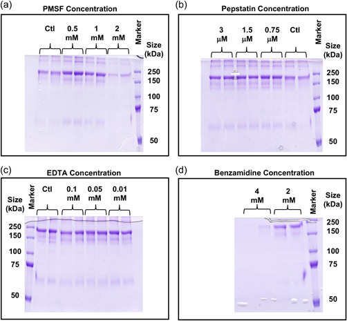
Mammalian cells, particularly Chinese hamster ovary cells, are the dominant system for the production of protein-based biotherapeutics, however, product degradation, particularly of Fc-fusion proteins, is sometimes observed that impacts the quality of the protein generated. Here, we identify the site of fragmentation of a model immunoglobulin G1 Fc-fusion protein, show that the observed clipping and aggregation are decreased by reduced temperature culturing, that the fragmentation/clipping is intracellular, and that reduced clipping at a lower temperature (<37°C) relates to mesenger RNA (mRNA) translation elongation. We subsequently show that reduced fragmentation can be achieved at 37°C by addition of chemical reagents that slow translation elongation. We then modified mRNA translation elongation speeds by designing different transcript sequences for the Fc-fusion protein based on alternative codon usage and improved the product yield at 37°C, and the ratio of intact to a fragmented product. Our data suggest that rapid elongation results in misfolding that decreases product fidelity, generating a region susceptible to degradation/proteolysis, whilst the slowing of mRNA translation improves the folding, reducing susceptibility to fragmentation. Manipulation of mRNA translation and/or the target Fc-fusion transcript is, therefore, an approach that can be applied to potentially reduce fragmentation of clipping-prone Fc-fusion proteins.

Keywords: Chinese hamster ovary (CHO) cells; Fc-fusion protein; clipping; fragmentation; mRNA translation elongation.


Conflict of interest statement

Atul Mohindra and Colin M. Jaques are employed by Lonza Biologics. Lonza Biologics owns and licenses the GS Gene Expression System®, CHOK1SV®, and CHOK1SV GS‐KO® cell lines. The remaining authors declare no conflict of interest.

Figures

Figure 1
Figure 1
Nonreducing SDS‐PAGE analysis of Protein A‐purified supernatant from two Lonza CHOK1SV® IgG1 Fc‐fusion protein‐expressing cell lines and subsequent determination of cleavage site (a) and 35S radiolabelling and pulse‐chase analysis of fusion product species produced from a stable CHO cell line by nonreduced SDS‐PAGE followed by radioautography. (a) Nonreducing SDS‐PAGE analysis of Protein A‐purified material from two IgG1 Fc‐fusion protein expressing CHOK1SV® cell lines and the different species present indicated. The intact product (indicated as intact dimer), single chain fragmented (indicated as fusion/fragmented product dimer) and doubly fragmented (fragment dimer) products were excised from the gel and analyzed by N‐terminal sequencing. This analysis showed cleavage/clipping to occur between an R (Arg) and S (Ser) residue to yield a protein lacking the “active” N‐terminal fusion part of the molecule. (b) Supernatant from stably producing cultures spiked with 35S radiolabel to monitor protein synthesis. Protein was precipitated using ice‐cold acetone. Lanes 1 and 2, supernatant precipitated at 20 h after addition of the 35S radiolabel to spent media with cells seeded at 0.3 × 106 cells/ml; Lane 3, supernatant precipitated at 44 h after addition of the 35S radiolabel (24 h after pulse chase) to spent media with cells seeded at 0.3 × 106 cells/ml. (c) Cell lysate samples from cultures spiked with 35S radiolabel to monitor protein synthesis. Samples were immunoprecipitated with antifusion section antibody followed by Protein A Sepharose bead pull‐down. Lane 1, cell lysate at 20 h after addition of the 35S radiolabel to spent media with cells seeded at 0.3 × 106 cells/ml; Lane 2, cell lysate at 44 h after addition of the 35S radiolabel (24 h after pulse chase) to spent media with cells seeded at 0.3 × 106 cells/ml. In (b) and (c), because the marker proteins are not radioactive, their bands were marked onto the film based on their positions clearly visible on the blot below the film. CHO, Chinese hamster ovary; IgG, immunoglobulin G; SDS‐PAGE, sodium dodecyl sulphate‐polyacrylamide gel electrophoresis.
Figure 2
Figure 2
Addition of protease inhibitors to a stable CHOK1SV® producing IgG1 HC Fc‐fusion cell line and the impact on fragmentation. Various classes of protease inhibitor were incubated with a cell line stably expressing the model IgG1 HC Fc‐fusion protein for 6 days in bath culture, adding the concentration of inhibitor indicated at Day 0 and again after 3 days. Supernatant samples were harvested after 6 days of culture, and the product purified using Protein A chromatography and analyzed by nonreducing SDS‐PAGE. (a) Addition of 0.5, 1, and 2 mM PMSF or ethanol control. (b) addition of 0.75, 1.5, and 3 µM pepstatin or ethanol‐treated control. (c) addition of 0.01, 0.05, and 0.1 mM EDTA or control. (d) addition of 2 and 4 mM benzamidine. Staining was performed with Coomassie brilliant blue G250. CHO, Chinese hamster ovary; PMSF, phenylmethanesulfonyl fluoride; SDS‐PAGE, sodium dodecyl sulphate‐polyacrylamide gel electrophoresis.
Figure 3
Figure 3
A temperature shift to 32°C during culture reduces IgG1 HC Fc‐fusion molecule fragmentation levels. (a) Supernatant from IgG1 HC Fc‐fusion stably expressing cell lines A and B were cultured at either 37 or 32°C for 7 days. At the end of incubation, supernatant from the cultures was harvested, purified by Protein A chromatography, and analyzed by nonreducing SDS‐PAGE followed by visualization of the bands present by Coomassie blue staining. The image shows supernatant samples from Day 7 of culture. (b) SDS‐PAGE analysis of the Protein A‐purified product from cell line A after a 7 day batch culture at 32°C. (c) Supernatant samples from HC Fc‐fusion stably expressing cell line A cultured at either 32°C or 37°C. Supernatant was harvested on Day 4 of culture and analyzed by nonreducing SDS‐PAGE followed by western blot analysis. Western blots were probed with anti‐HC antibody. (d) Densitometry analysis of the average band intensity of the intact or fragment bands at 37 or 32°C in (b). (e) The intact to fragment band intensity ratio calculated from the data in (d). IgG, immunoglobulin G; SDS‐PAGE, sodium dodecyl sulphate‐polyacrylamide gel electrophoresis.
Figure 4
Figure 4
Slowing of translation elongation by the addition of the chemical inhibitor cycloheximide to cultures of CHOK1SV cells stably expressing a model IgG1 Fc‐fusion molecule results in reduced fragmentation. Cultures of the cell line A stably expressing the fusion protein, incubated with (a) 0 (control), 0.05, 0.025 and (b) 0.05, 0.01, and 0.005 µg/ml cycloheximide. All concentrations were investigated in triplicate. Cultures were harvested after 7 days and the supernatant was purified using Protein A chromatography, the elution fractions were concentrated and analyzed on an 8% nonreduced SDS‐PAGE with Coomassie staining. (c, d) Densitometry analysis of the average band intensity of the intact or fragment bands in (a, b). (e) The intact to fragment band intensity ratio calculated from the data in (c, d) (note as there is no fragment detected at 0.05 µg/ml cycloheximide there is no data for this concentration). IgG, immunoglobulin G; SDS‐PAGE, sodium dodecyl sulphate‐polyacrylamide gel electrophoresis.
Figure 5
Figure 5
The impact on Fc‐fusion protein fragmentation via direct manipulation of eEF2 amounts using shRNA. Stably IgG1 Fc‐fusion protein‐producing cell line A and the host cell line were transfected with shRNAs designed to the CHO cell genome eEF2 sequence using the GeneCLIP system. Supernatant and cell lysate samples were taken 24 and 48 h after transfection. Western blot analysis was performed using an anti‐eEF2 antibody (Cell Signalling) with β‐actin as a loading control. Supernatant was analyzed by western blot analysis using an anti‐HC antibody (a). (b) Densitometry analysis was performed using ImageJ software to confirm eEF2 knockdown. (c) Densitometry analysis of the average band intensity of the intact or fragment bands, as indicated in (a) for the 48 h time point and the calculated intact to fragment band ratio. CHO, Chinese hamster ovary; eEF2, elongation factor 2; IgG, immunoglobulin G; shRNA, short hairpin RNA.
Figure 6
Figure 6
Determining the impact on fragmentation of manipulation of translation rates via messenger RNA sequence. The CHO host cell line was transiently transfected using electroporation with sequences designed via standard commercial codon optimization (standard) or using a balance of fast (fast) or deoptimized (slow) codons in the translation model developed by Chu et al. (2014) and using Chinese hamster tRNA abundances. Supernatant samples were harvested 96 h after transfection and assessed for recombinant product levels via western blot analysis using an anti‐HC antibody (Sigma‐Aldrich). CHO, Chinese hamster ovary; tRNA, transfer RNA.
Figure 7
Figure 7
Proposed hypothesis for the reduced fragmentation of the immunoglobulin G1 Fc‐fusion molecule upon slowed messenger RNA translation elongation

Similar articles

Cited by

References

    1. Bryan, L. , Henry, H. , Barron, N. , Gallagher, C. , Kelly, R. M. , Frye, C. C. , Osborne, M. D. , Clynes, M. , Meleady, P. (2021). Differential expression of miRNAs and functional role of miR‐200a in high and low productivity CHO cells expressing an Fc fusion protein, Biotechnology Letters, 43, 1551–1563. 10.1007/s10529-021-03153-7 - DOI - PMC - PubMed
    1. Budge, J. D. , Knight, T. J. , Povey, J. , Roobol, J. , Brown, I. R. , Singh, G. , Dean, A. , Turner, S. , Jaques, C. M. , Young, R. J. , Racher, A. J. , Smales, C. M. (2020). Engineering of Chinese hamster ovary cell lipid metabolism results in an expanded ER and enhanced recombinant biotherapeutic protein production, Metabolic Engineering, 57, 203–216. 10.1016/j.ymben.2019.11.007 - DOI - PMC - PubMed
    1. Budge, J. D. , Roobol, J. , Singh, G. , Mozzanino, T. , Knight, T. J. , Povy, J. , Dean, A. , Turner, S. J. , Jaques, C. M. , Young, R. J. , Racher, A. J. , Smales, C. M. (2021). A proline metabolism selection system and its application to the engineering of lipid biosynthesis in Chinese hamster ovary cells, Metabolic Engineering Communications, 13, e00179. 10.1016/j.mec.2021.e00179 - DOI - PMC - PubMed
    1. Chakrabarti, S. , Barrow C. J., Kanwar R. K., Ramana V., Kanwar J. R. (2016). ‘Studies to prevent degradation of recombinant Fc‐fusion protein expressed in mammalian cell line and protein characterization’, International Journal of Molecular Sciences, 17(6), 913. 10.3390/ijms17060913 - DOI - PMC - PubMed
    1. Chu, D. , Thompson, J. & von der Haar, T. (2014). Charting the dynamics of translation, Biosystems, 119, 1–9. 10.1016/j.biosystems.2014.02.005 - DOI - PubMed

Publication types

LinkOut - more resources