. 2022 Oct;48(4):183.
doi: 10.3892/or.2022.8398. Epub 2022 Sep 9.

High expression of Sam68 contributes to metastasis by regulating vimentin expression and a motile phenotype in oral squamous cell carcinoma

Affiliations

High expression of Sam68 contributes to metastasis by regulating vimentin expression and a motile phenotype in oral squamous cell carcinoma

Takuya Komiyama et al. Oncol Rep. 2022 Oct.

Abstract

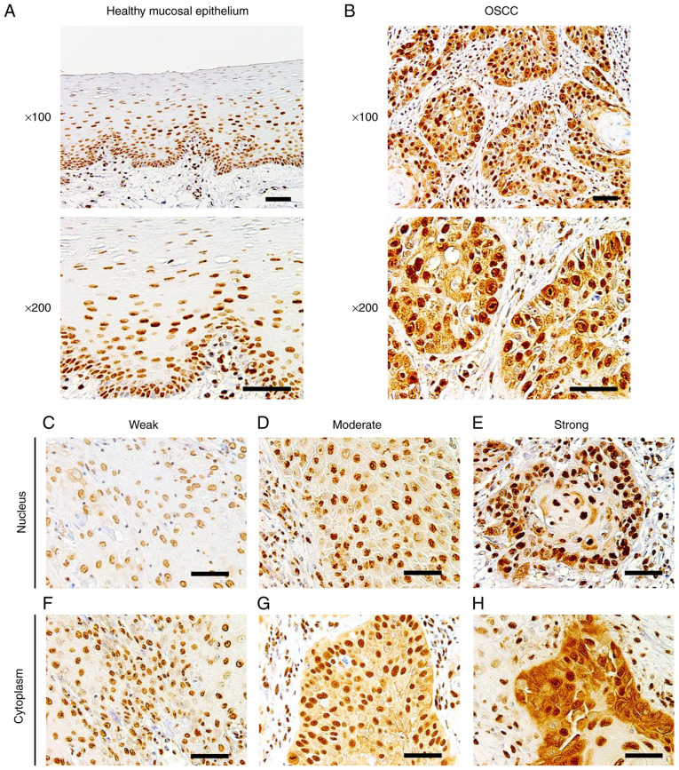
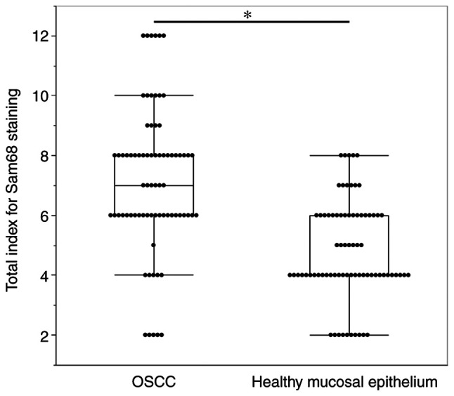
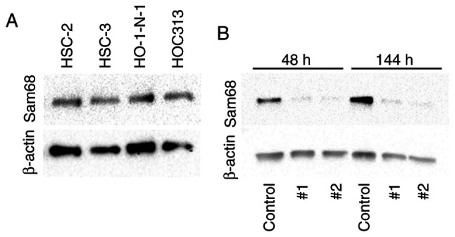
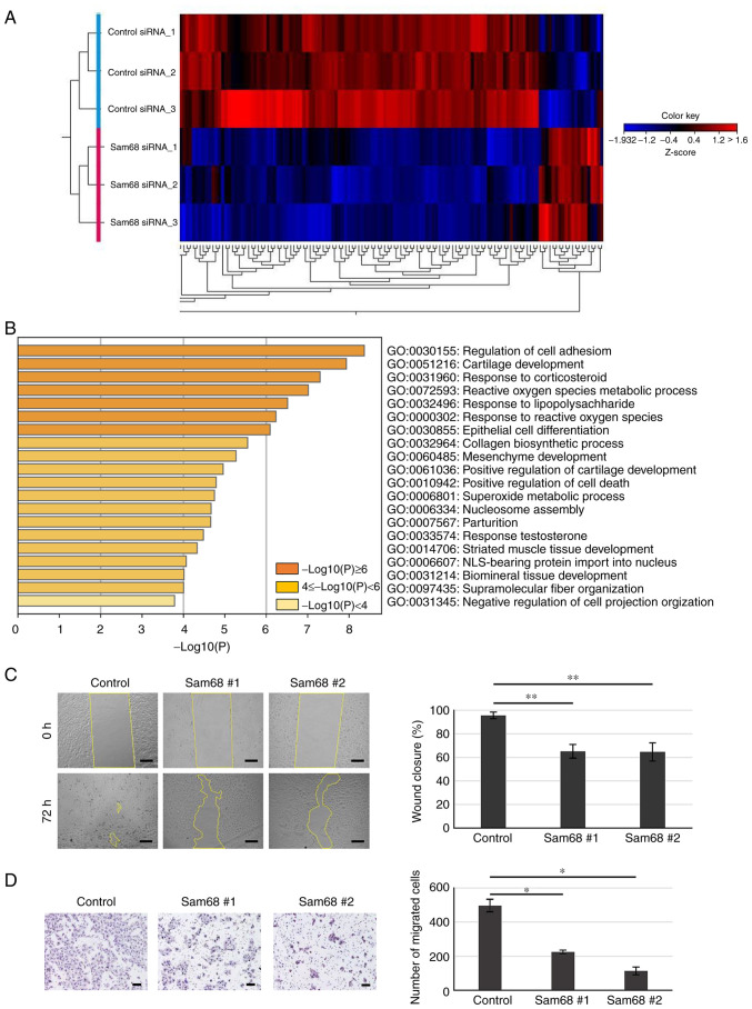
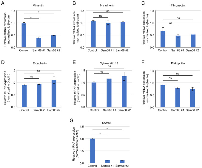
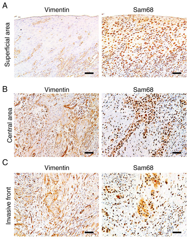
The present study aimed to investigate the clinical and biological significance of Src‑associated in mitosis 68 kDa (Sam68) in oral squamous cell carcinoma (OSCC). Immunohistochemical analysis was performed on tissue samples obtained from 77 patients with OSCC. Univariate analysis revealed that the high expression of Sam68 was significantly correlated with advanced pathological T stage (P=0.01), positive lymphovascular invasion (P=0.01), and pathological cervical lymph node metastasis (P<0.01). Moreover, multivariate analysis demonstrated that the high expression of Sam68 was an independent predictive factor for cervical lymph node metastasis (odds ratio, 4.39; 95% confidence interval, 1.49‑14.23; P<0.01). These results indicated that high Sam68 expression contributed to tumor progression, especially cervical lymph node metastasis, in OSCC. mRNA sequencing was also performed to assess the changes in the transcriptome between OSCC cells with Sam68 knockdown and control cells with the aim of elucidating the biological roles of Sam68. Gene Ontology enrichment analysis revealed that downregulated differentially expressed genes (DEGs) were concentrated in some biological processes related to epithelial‑mesenchymal transition. Among these DEGs, it was established that vimentin was particularly downregulated in these cells. It was also confirmed that Sam68 knockdown reduced the motility of OSCC cells. Furthermore, the immunohistochemical study of vimentin identified the association between vimentin expression and Sam68 expression as well as cervical lymph node metastasis. In conclusion, the present study suggested that the high expression of Sam68 may contribute to metastasis by regulating vimentin expression and a motile mesenchymal phenotype in OSCC.

Keywords: Src‑associated in mitosis 68 kDa; epithelial‑mesenchymal transition; metastasis; motility; oral cancer; vimentin.


Conflict of interest statement

The authors declare that they have no competing interests.

Figures

Figure 1.
Figure 1.
Immunohistochemistry of Sam68. (A) Representative images of healthy mucosal epithelium adjacent to OSCC, in which Sam68 expression was predominantly detected in the nucleus and negatively observed in the cytoplasm (magnification, ×100 and ×200; scale bar, 50 µm). (B) Representative image of OSCC, in which Sam68 expression was detected both in the nucleus and cytoplasm (magnification, ×100 and ×200; scale bar, 50 µm). (C-E) Nuclear expression of Sam68 with (C) weak, (D) moderate, and (E) strong staining intensity in OSCC cells (magnification, ×200; scale bar, 50 µm). (F-H) Cytoplasmic expression of Sam68 with (C) weak, (D) moderate, and (E) strong staining intensity in OSCC tissue (magnification, ×200; scale bar, 50 µm). Sam68, Src-associated in mitosis 68 kDa; OSCC, oral squamous cell carcinoma.
Figure 2.
Figure 2.
Comparison of the total staining index of Sam68 between OSCC cells and adjacent healthy mucosal epithelium. Mann-Whitney U test. *P<0.01. Sam68, Src-associated in mitosis 68 kDa; OSCC, oral squamous cell carcinoma.
Figure 3.
Figure 3.
Expression of Sam68 in oral cancer cell lines. (A) Western blot analysis of Sam68 in oral cancer cells (HSC2, HSC3, HO-1-N-1, and HOC313). (B) Sam68 siRNA (#1 or #2) or control siRNA (control) was transfected into HO-1-N-1 cells, and the proteins were estimated at 48 or 144 h by western blotting. Sam68, Src-associated in mitosis 68 kDa.
Figure 4.
Figure 4.
Inhibition of migration activity in Sam68-knockdown OSCC cells. (A) Heat map representation of 150 DEGs identified in mRNA sequencing between Sam68 siRNA #1-transfected HO-1-N-1 cells (n=3) and control siRNA-transfected cells (n=3). (B) GO enrichment analysis of 130 downregulated DEGs in Sam68-knockdown HO-1-N-1 cells. Bar graph of enriched GO terms (biological process) across input gene lists, colored by P-values. (C) HO-1-N-1 cells transfected with control siRNA (control) or Sam68 siRNA (Sam68 #1 or #2) were analyzed for wound closure at 72 h after wounding. Scale bar, 200 µm. Data are expressed as the mean ± SD. Dunnett's test compared with the control. **P<0.05. (D) HO-1-N-1 cells transfected with control siRNA (control) or Sam68 siRNA (Sam68 #1 or #2) were cultured in a Transwell chamber for 48 h. Migrated cells were stained and counted. Scale bar, 200 µm. Data are expressed as the mean ± SD. Dunnett's test compared with the control. *P<0.01. Sam68, Src-associated in mitosis 68 kDa; OSCC, oral squamous cell carcinoma; DEGs, differentially expressed genes; GO, Gene Ontology; siRNA, small interfering RNA.
Figure 5.
Figure 5.
Validation of gene expression by RT-qPCR. (A and G) Comparison of mRNA expression of DEGs and (B-F) non-DEGs associated with the epithelial-mesenchymal transition between HO-1-N-1 cells transfected with control siRNA (control) or Sam68 siRNA (Sam68 #1 or #2). All data are expressed as the mean ± SD. Dunnett's test compared with the control. *P<0.01. ns, not significant. RT-qPCR, reverse transcription-quantitative PCR; DEGs, differentially expressed genes; siRNA, small interfering RNA; Sam68, Src-associated in mitosis 68 kDa; ns, not significant.
Figure 6.
Figure 6.
Immunohistochemical staining showing (A) negative and (C) positive expression of vimentin and (B and D) hematoxylin and eosin staining (magnification, ×100; scale bar, 50 µm).
Figure 7.
Figure 7.
Immunohistochemistry of vimentin and Sam68 in the same tumor. (A) Representative image of the broad sheet of tumor cells without vimentin expression at the superficial area. (B) Representative image of the small cords of tumor cells exhibiting positive vimentin expression and higher Sam68 immunoreactivity at the central area. (C) Representative image of the small cords and clusters of tumor cells exhibiting positive vimentin expression and higher Sam68 immunoreactivity at the invasive front. (magnification, ×100; scale bar, 50 µm). Sam68, Src-associated in mitosis 68 kDa.

Similar articles

Cited by

References

    1. Bray F, Ferlay J, Soerjomataram I, Siegel RL, Torre LA, Jemal A. Global cancer statistics 2018: GLOBOCAN estimates of incidence and mortality worldwide for 36 cancers in 185 countries. CA Cancer J Clin. 2018;68:394–424. doi: 10.3322/caac.21492. - DOI - PubMed
    1. El-Naggar AK, Chan JKC, Grandis JR, Takata T, Slootweg PJ. 4th edition. IARC Press; Lyon: 2017. WHO classification of head and neck tumours.
    1. Sato J, Kitagawa Y, Watanabe S, Asaka T, Ohga N, Hirata K, Shiga T, Satoh A, Tamaki N. Hypoxic volume evaluated by 18F-fluoromisonidazole positron emission tomography (FMISO-PET) may be a prognostic factor in patients with oral squamous cell carcinoma: Preliminary analyses. Int J Oral Maxillofac Surg. 2018;47:553–560. doi: 10.1016/j.ijom.2017.09.007. - DOI - PubMed
    1. Larsen SR, Johansen J, Sørensen JA, Krogdahl A. The prognostic significance of histological features in oral squamous cell carcinoma. J Oral Pathol Med. 2009;38:657–662. doi: 10.1111/j.1600-0714.2009.00797.x. - DOI - PubMed
    1. Oikawa Y, Kugimoto T, Kashima Y, Okuyama K, Ohsako T, Kuroshima T, Hirai H, Tomioka H, Shimamoto H, Michi Y, Harada H. Surgical treatment for oral tongue squamous cell carcinoma: A retrospective study of 432 patients. Glob Health Med. 2021;3:157–162. doi: 10.35772/ghm.2020.01084. - DOI - PMC - PubMed

MeSH terms