The decoupling of structural and functional connectivity of auditory networks in individuals at clinical high-risk for psychosis
- PMID: 36083108
- PMCID: PMC10399965
- DOI: 10.1080/15622975.2022.2112974
The decoupling of structural and functional connectivity of auditory networks in individuals at clinical high-risk for psychosis
Abstract
Objectives: Disrupted auditory networks play an important role in the pathophysiology of psychosis, with abnormalities already observed in individuals at clinical high-risk for psychosis (CHR). Here, we examine structural and functional connectivity of an auditory network in CHR utilising state-of-the-art electroencephalography and diffusion imaging techniques.
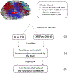
Methods: Twenty-six CHR subjects and 13 healthy controls (HC) underwent diffusion MRI and electroencephalography while performing an auditory task. We investigated structural connectivity, measured as fractional anisotropy in the Arcuate Fasciculus (AF), Cingulum Bundle, and Superior Longitudinal Fasciculus-II. Gamma-band lagged-phase synchronisation, a functional connectivity measure, was calculated between cortical regions connected by these tracts.
Results: CHR subjects showed significantly higher structural connectivity in the right AF than HC (p < .001). Although non-significant, functional connectivity between cortical areas connected by the AF was lower in CHR than HC (p = .078). Structural and functional connectivity were correlated in HC (p = .056) but not in CHR (p = .29).
Conclusions: We observe significant differences in structural connectivity of the AF, without a concomitant significant change in functional connectivity in CHR subjects. This may suggest that the CHR state is characterised by a decoupling of structural and functional connectivity, possibly due to abnormal white matter maturation.
Keywords: EEG; MRI; biological psychiatry; imaging; schizophrenia.
Conflict of interest statement
Declaration of Interest
The authors have declared that there are no conflicts of interest in relation to the subject of this study.
Figures


References
-
- Assaf Y, Pasternak O. 2008. Diffusion Tensor Imaging (DTI)-based White Matter Mapping in Brain Research: A Review. J Mol Neurosci. 34(1):51–61. - PubMed
-
- Billah T, Bouix S, Rathi Y. 2019. Various MRI Conversion Tools. https://githubcom/pnlbwh/conversion.
-
- Bloemen OJN, de Koning MB, Schmitz N, Nieman DH, Becker HE, de Haan L, Dingemans P, Linszen DH, van Amelsvoort TAMJ. 2010. White-matter markers for psychosis in a prospective ultra-high-risk cohort. Psychol Med. 40(8):1297–1304. - PubMed
Publication types
MeSH terms
Grants and funding
- R01 MH102377/MH/NIMH NIH HHS/United States
- K24 MH110807/MH/NIMH NIH HHS/United States
- P41 EB015898/EB/NIBIB NIH HHS/United States
- R03 MH110745/MH/NIMH NIH HHS/United States
- R01 MH119222/MH/NIMH NIH HHS/United States
- R01 MH074794/MH/NIMH NIH HHS/United States
- P41 EB028741/EB/NIBIB NIH HHS/United States
- R01 MH108574/MH/NIMH NIH HHS/United States
- K01 MH115247/MH/NIMH NIH HHS/United States
- U01 CA199459/CA/NCI NIH HHS/United States
- R01 NS125781/NS/NINDS NIH HHS/United States
- U01 MH109977/MH/NIMH NIH HHS/United States
- P41 EB015902/EB/NIBIB NIH HHS/United States
LinkOut - more resources
Full Text Sources
Other Literature Sources
Medical
