. 2022 Nov;24(11):2351-2366.
doi: 10.1016/j.gim.2022.08.006. Epub 2022 Sep 9.

Genomic and phenotypic characterization of 404 individuals with neurodevelopmental disorders caused by CTNNB1 variants

Sayaka Kayumi  1 Luis A Pérez-Jurado  2 María Palomares  3 Sneha Rangu  4 Sarah E Sheppard  5 Wendy K Chung  6 Michael C Kruer  7 Mira Kharbanda  8 David J Amor  9 George McGillivray  10 Julie S Cohen  11 Sixto García-Miñaúr  3 Clare L van Eyk  1 Kelly Harper  1 Lachlan A Jolly  12 Dani L Webber  1 Christopher P Barnett  13 Fernando Santos-Simarro  3 Marta Pacio-Míguez  3 Angela Del Pozo  3 Somayeh Bakhtiari  7 Matthew Deardorff  14 Holly A Dubbs  15 Kosuke Izumi  16 Katheryn Grand  17 Christopher Gray  18 Paul R Mark  19 Elizabeth J Bhoj  20 Dong Li  21 Xilma R Ortiz-Gonzalez  22 Beth Keena  23 Elaine H Zackai  20 Ethan M Goldberg  24 Guiomar Perez de Nanclares  25 Arrate Pereda  25 Isabel Llano-Rivas  26 Ignacio Arroyo  27 María Ángeles Fernández-Cuesta  28 Christel Thauvin-Robinet  29 Laurence Faivre  30 Aurore Garde  31 Benoit Mazel  31 Ange-Line Bruel  32 Michael L Tress  33 Eva Brilstra  34 Amena Smith Fine  11 Kylie E Crompton  9 Alexander P A Stegmann  35 Margje Sinnema  35 Servi C J Stevens  35 Joost Nicolai  36 Gaetan Lesca  37 Laurence Lion-François  38 Damien Haye  37 Nicolas Chatron  37 Amelie Piton  39 Mathilde Nizon  40 Benjamin Cogne  40 Siddharth Srivastava  41 Jennifer Bassetti  42 Candace Muss  43 Karen W Gripp  43 Rebecca A Procopio  44 Francisca Millan  45 Michelle M Morrow  45 Melissa Assaf  46 Andres Moreno-De-Luca  47 Shelagh Joss  48 Mark J Hamilton  48 Marta Bertoli  49 Nicola Foulds  8 Shane McKee  50 Alastair H MacLennan  1 Jozef Gecz  51 Mark A Corbett  52
Affiliations

Genomic and phenotypic characterization of 404 individuals with neurodevelopmental disorders caused by CTNNB1 variants

Sayaka Kayumi et al. Genet Med. 2022 Nov.

Abstract

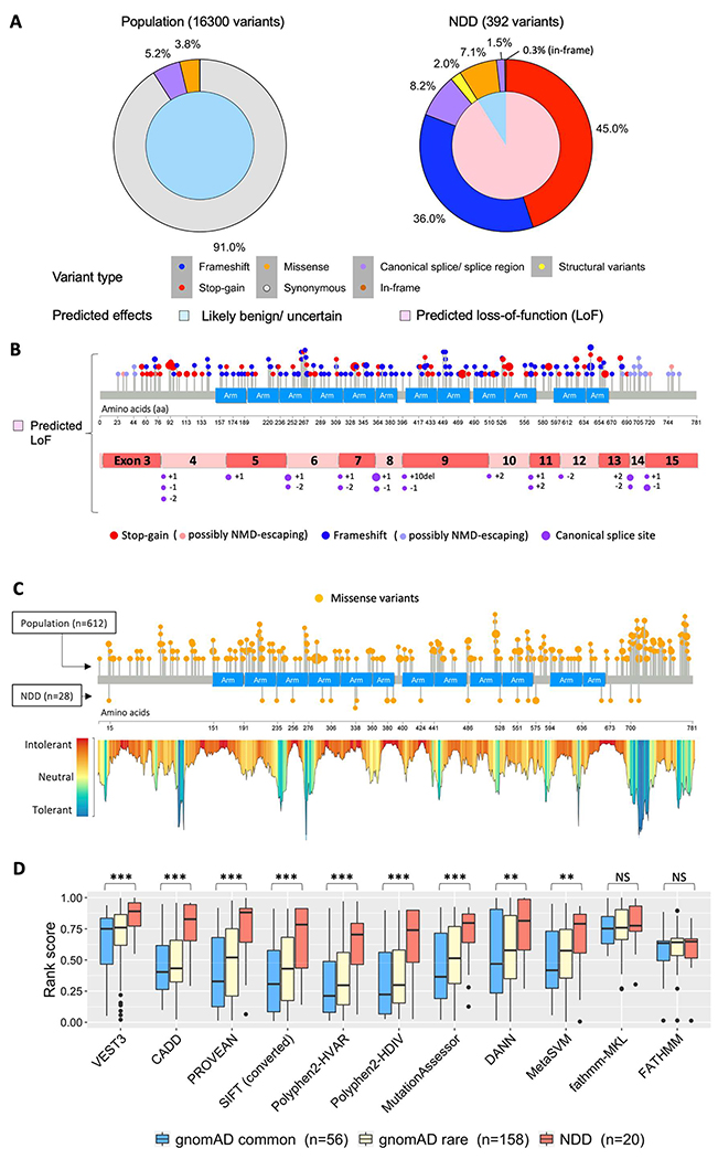
Purpose: Germline loss-of-function variants in CTNNB1 cause neurodevelopmental disorder with spastic diplegia and visual defects (NEDSDV; OMIM 615075) and are the most frequent, recurrent monogenic cause of cerebral palsy (CP). We investigated the range of clinical phenotypes owing to disruptions of CTNNB1 to determine the association between NEDSDV and CP.

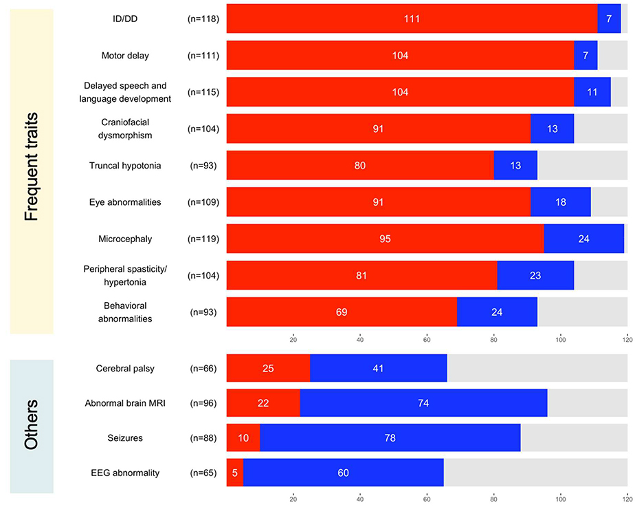
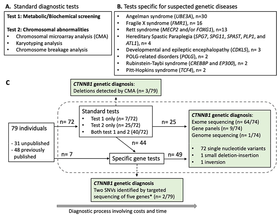
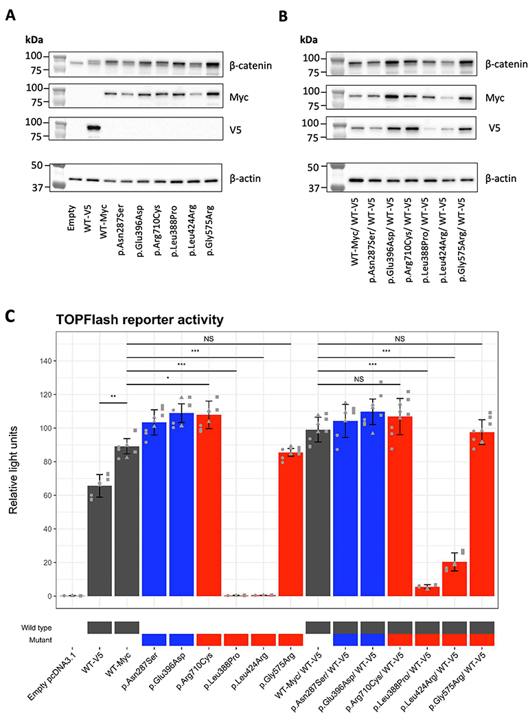
Methods: Genetic information from 404 individuals with collectively 392 pathogenic CTNNB1 variants were ascertained for the study. From these, detailed phenotypes for 52 previously unpublished individuals were collected and combined with 68 previously published individuals with comparable clinical information. The functional effects of selected CTNNB1 missense variants were assessed using TOPFlash assay.

Results: The phenotypes associated with pathogenic CTNNB1 variants were similar. A diagnosis of CP was not significantly associated with any set of traits that defined a specific phenotypic subgroup, indicating that CP is not additional to NEDSDV. Two CTNNB1 missense variants were dominant negative regulators of WNT signaling, highlighting the utility of the TOPFlash assay to functionally assess variants.

Conclusion: NEDSDV is a clinically homogeneous disorder irrespective of initial clinical diagnoses, including CP, or entry points for genetic testing.

Keywords: Autism; Cerebral palsy; Familial exudative vitreoretinopathy; Microcephaly; Wnt beta catenin signaling pathway.


Conflict of interest statement

Conflict of Interest F.M. and M.M.M. are employees of GeneDX, Inc. All other authors declare no conflict of interest.

Figures

Figure 1.
Figure 1.
Graphical summary of 52 unpublished individuals with pathogenic variants in CTNNB1. (A) The division of males and females in the cohort. (B) The proportions of different types of variants. Colors represent different variant types: stop-gain variant (red), frameshift variant (blue), splice donor variant (purple), structural variant (yellow), and missense variant (orange). (C) Lollipop plot shows β-catenin structure at the bottom and blue boxes represent Armadillo repeat domains in β-catenin. Each dot represents a CTNNB1 variant identified in unpublished individuals. Recurrent variants identified in two or more unrelated individuals are labelled with the amino acid changes with the number of individuals in brackets. (D) Variants affecting splice donor sites were identified in two individuals. (E) Structural variants of a deletion and a balanced inversion in chromosome 3 were identified in two individuals. (F) Facial images of 10 individuals with CTNNB1 variants. Images were collected from unpublished individual 9, 10, 19 (at the age of four years old and nine years old), 28, 29, 30, 34 (at the age of six years old and 16 years and 9 months old), 40, 44, and previously published individual 180.
Figure 2.
Figure 2.
Neurological traits associated with pathogenic or likely pathogenic variants in CTNNB1. Traits that were frequently identified in a cohort of unpublished and previously published individuals are summarized at the top. Other relevant traits discussed in the present study are summarized at the bottom. Bar charts show the number of affected (red), unaffected (blue), and unknown (grey) individuals per trait. The number of individuals known for their affected status per trait is shown in brackets next to each trait. ID, intellectual disability; DD, developmental delay.
Figure 3.
Figure 3.
Diagnostic pathways of 79 individuals prior to their CTNNB1 genetic diagnosis. (A) Standard diagnostic tests performed during diagnostic process. (B) A list of tests to assess suspected, specific genetic diseases that were performed in two or more individuals. (C) A graphical summary of diagnostic pathways of 79 individuals prior to their CTNNB1 genetic diagnosis. Seventy-four single nucleotide variants (SNVs) in CTNNB1 were identified through exome sequencing, targeted next-generation sequencing panels, or genome sequencing. These variants included 31 frameshift, 29 stop-gain, eight canonical splice site, and four missense variants. Two variants (*) were exceptionally identified by targeted sequencing of five intellectual disability genes including CTNNB1.
Figure 4.
Figure 4.
Analysis of phenotypic outcome by CTNNB1 variants type. (A) Distinct patterns of CTNNB1 variants identified in the general population and neurodevelopmental disorders (NDD) with different predicted effects on CTNNB1. Inner pie charts show the ratio of variants with predicted effects of likely benign or uncertain (light blue) and loss of function (pink). Outer pie charts show the percentage of variants by type: synonymous (grey), missense (orange), frameshift (blue), stop-gain (red), splicing site (purple), in-frame insertions / deletions (brown), and structural variants mainly with deletions (yellow). Percentage labels of variant types are shown when the values are larger than 0.1%. (B) Variant plots showing distribution of stop-gain, frameshift, and canonical splice site variants of CTNNB1 identified in individuals with NDD. Lollipop plot shows μ-catenin structure at the bottom and blue box represents Armadillo repeat domains. Each dot represents an individual with stop-gain (red) or frameshift (blue) variant of CTNNB1. The larger size of a dot indicates multiple individuals with the same variant. Variants predicted to escape nonsense-mediated mRNA decay (NMD) are indicated with lighter blue (frameshift) and lighter red (stop-gain). CTNNB1 mRNA structure shows exons with canonical splice site variants (purple) likely affecting normal splicing of CTNNB1. Locations of these splice site variants in intron regions were noted with the number of nucleotides from the last nucleotide of an exon (+) or the first nucleotide of an exon (−). (C) Analysis of missense CTNNB1 variants identified in individuals with NDD compared to likely benign variants identified in the general population. Lollipop plots show distribution of missense variants of CTNNB1 (orange) identified in the general population (above) or individuals with NDD (below). Landscape of CTNNB1 variant tolerance generated using MetaDome is shown under the lollipop plot. (D) Summary of deleterious predictions of missense CTNNB1 variants using 11 predictive tools. Box plots show 1st quartile (bottom) to 3rd quartile (top) with each median value at the center. SIFT scores were calculated as 1-SIFT raw score. Student’s t-test was applied to assess the difference of NDD variants against population variants with allele frequency equal or greater than 1.0 × 10E-05 (gnomAD common). The significance marked with “***”=0.001,“**”=0.01,“*”=0.05, or “NS”=Not significant.
Figure 5.
Figure 5.
Functional assessment of missense CTNNB1 variants using TOPFlash assay. (A-B) Detection of Myc or V5-tagged wild-type (WT) β-catenin proteins and Myc-tagged mutant β-catenin proteins transfected into HEK293T cells by western blot. Expression constructs were transfected without co-transfection (A) and with co-transfection of a V5-tagged wild-type β-catenin (B). Molecular sizes of standard protein markers were indicated on the left of blots. Endogenous and exogenous β-catenin were detected with a β-catenin antibody (amino acid 571–781). Exogenous β-catenin was identified with a V5 antibody, and a Myc antibody. Endogenous levels of β-actin were detected to show equal loading by western blot. Full blots are available in Supplementary Figure 5. (C) Effects of missense CTNNB1 variants Wnt signaling as measured by the TOPFlash assay. Relative luciferase activity measured using the TOPFlash assay in HEK293T cells transfected with expression vectors for wild-type β-catenin or mutant β-catenin or an equal mix with wildtype β-catenin tagged with V5. Wild type, gnomAD variants, and pathogenic/likely pathogenic variants are highlighted in grey, blue, and red on the X-axis labels, respectively. Assay was performed in triplicate (shown with different shaped data points) with three technical replicate samples for each assay. Error bars indicate standard deviations between the three independent experiments. Student’s t-test was applied to assess the difference of relative light unit of pathogenic/likely pathogenic variants against that of wild-type β-catenin. The significance marked with “***”=0.001,“**”=0.01,“*”=0.05, or “NS”=Not significant.

References

    1. Vissers LELM, van Nimwegen KJM, Schieving JH, et al. A clinical utility study of exome sequencing versus conventional genetic testing in pediatric neurology. Genet Med. 2017;19(9):1055–1063. doi:10.1038/gim.2017.1 - DOI - PMC - PubMed
    1. Chopra M, Gable DL, Love- Nichols J, et al. Mendelian etiologies identified with whole exome sequencing in cerebral palsy. Ann Clin Transl Neurol. 2022;9(2):193–205. doi:10.1002/acn3.51506 - DOI - PMC - PubMed
    1. van Eyk CL, Webber DL, Minoche AE, et al. Yield of clinically reportable genetic variants in unselected cerebral palsy by whole genome sequencing. Npj Genomic Med. 2021;6(1):74. doi:10.1038/s41525-021-00238-0 - DOI - PMC - PubMed
    1. Moreno-De-Luca A, Millan F, Pesacreta DR, et al. Molecular Diagnostic Yield of Exome Sequencing in Patients With Cerebral Palsy. JAMA. 2021;325(5):467. doi:10.1001/jama.2020.26148 - DOI - PMC - PubMed
    1. Kuechler A, Willemsen MH, Albrecht B, et al. De novo mutations in beta-catenin (CTNNB1) appear to be a frequent cause of intellectual disability: expanding the mutational and clinical spectrum. Hum Genet. 2015;134(1):97–109. doi:10.1007/s00439-014-1498-1 - DOI - PubMed

Publication types

LinkOut - more resources