. 2022 Nov;64(11):2191-2201.
doi: 10.1007/s00234-022-03047-z. Epub 2022 Sep 9.

The prevalence of redundant nerve roots in standing positional MRI decreases by half in supine and almost to zero in flexed seated position: a retrospective cross-sectional cohort study

Affiliations

The prevalence of redundant nerve roots in standing positional MRI decreases by half in supine and almost to zero in flexed seated position: a retrospective cross-sectional cohort study

Luca Papavero et al. Neuroradiology. 2022 Nov.

Abstract

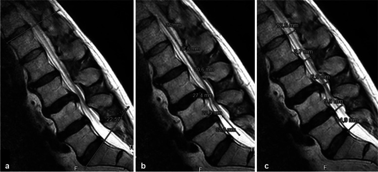
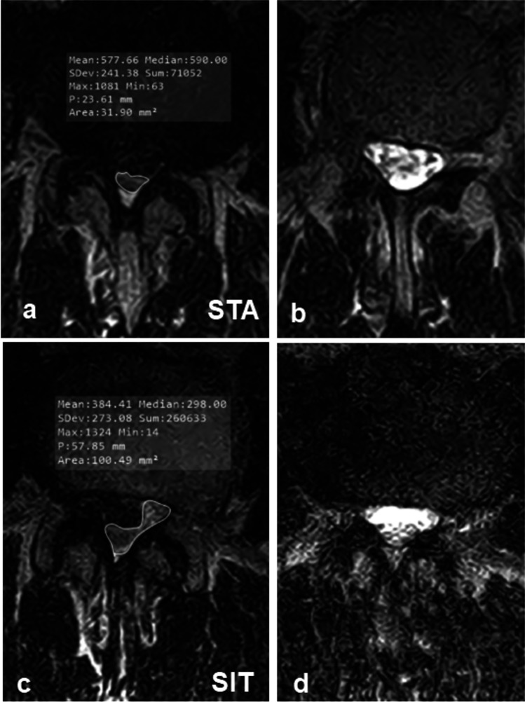
Purpose: This retrospective cross-sectional cohort study investigated the influence of posture on lordosis (LL), length of the spinal canal (LSC), anteroposterior diameter (APD L1-L5), dural cross-sectional area (DCSA) of the lumbar spinal canal, and the prevalence of redundant nerve roots (RNR) using positional magnetic resonance imaging (MRI) (0.6 T).

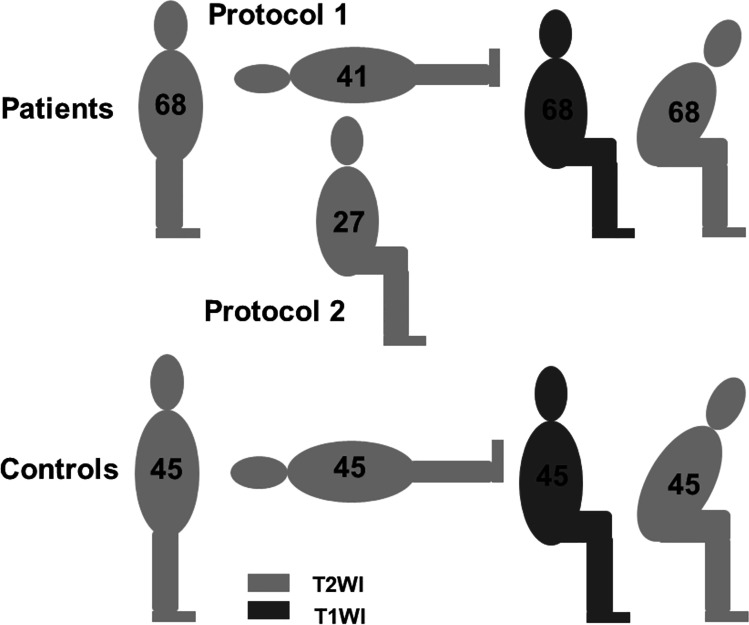
Methods: Sixty-eight patients with single-level degenerative central lumbar spinal stenosis (cLSS) presenting with RNR in the standing position (STA) were also investigated in supine (SUP) or neutral seated (SIT) and flexed seated (FLEX) positions. Additionally, 45 patients complaining of back pain and without MRI evidence of LSS were evaluated. Statistical significance was set at p < 0.05.

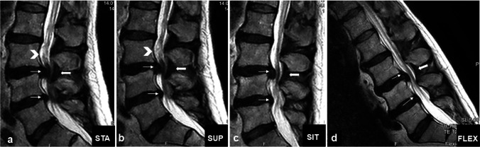
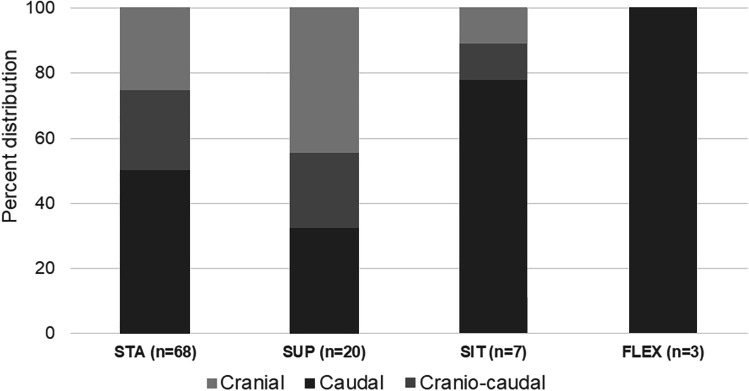
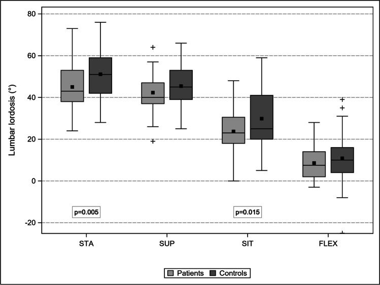
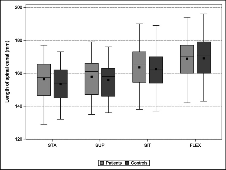
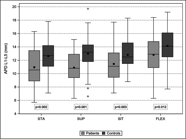
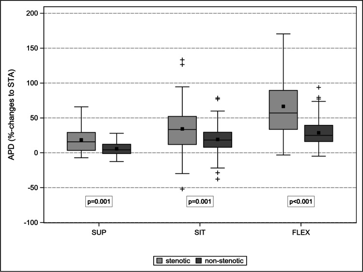
Results: Controls (A) and patients with cLSS (B) were comparable in terms of mean age (p = 0.88) and sex (p = 0.22). The progressive transition from STA to FLEX led to a comparable decrease in LL (p = 0.97), an increase in LSC (p = 0.80), and an increase in APD L1-L5 (p = 0.78). The APD of the stenotic level increased disproportionally between the different postures, up to 67% in FLEX compared to 29% in adjacent non-stenotic levels (p < 0.001). Therefore, the prevalence of RNR decreased to 49, 26, and 4% in SUP, SIT, and FLEX, respectively.

Conclusion: The prevalence of RNR in standing position was underestimated by half in supine position. Body postures modified LL, LSC, and APD similarly in patients and controls. Stenotic levels compensated for insufficient intraspinal volume with a disproportionate enlargement when switching from the STA to FLEX.

Keywords: Lumbar spine; Positional magnetic resonance imaging; Posture; Redundant nerve roots; Spinal stenosis.


Conflict of interest statement

We declare that we have no conflict of interest.

Figures

Fig. 1
Fig. 1
(Top) Study protocols of patients and (bottom) of controls
Fig. 2
Fig. 2
Parameters: a lumbar lordosis (LL): angle between the upper endplate L1 and the upper endplate S1. b Length of the spinal canal (LSC): sum of the segmental lengths parallel to the main bundle of cauda nerve roots between the upper endplate L1 and the upper endplate S1. c Anteroposterior diameter (APD): segmental sagittal diameter of the dural sac at the mid-disk level
Fig. 3
Fig. 3
Clinical case: a 73-year-old man presented with neurogenic claudication. In the previous 3 weeks, he experienced exacerbation of leg pain and new hypesthesia in the lower limbs. a Standing: RNR (arrowhead) cranial to the pincer stenosis caused by the buckling of disks (slim arrows) and yellow ligament (thick arrow). b No relevant difference in supine posture was observed. c In neutral sitting partial flattening of disks and yellow ligament with increase in the anteroposterior diameters and resolution of RNR was observed. d Enlargement of the stenotic level and complete resolution of the RNR
Fig. 4
Fig. 4
Same patient as in Fig. 1: dural cross-sectional area (DCSA) in standing position at the stenotic level (a) and 10-mm cranial (b) coiled and loop-shaped cauda nerve roots with positive sedimentation sign. c In sitting DCSA trebled, cauda nerve roots run perpendicular to the axial plane and the sedimentation sign became negative
Fig. 5
Fig. 5
Percent distribution of RNR location to the stenotic level. Although the data of some subgroups is small, there is a trend for caudal location of RNR in body postures influenced by axial gravity
Fig. 6
Fig. 6
Comparison of the change of LL between patients and controls: progressive flattening from STA to FLEX in both cohorts. The difference is significant only in STA and SIT
Fig. 7
Fig. 7
Comparison of lumbar spinal canal length (mm) changes between patients and controls: progressive and comparable lengthening from STA to FLEX
Fig. 8
Fig. 8
Comparison of the APD L1-L5 (mm) between patients and controls: the dynamic is comparable although the absolute values differ significantly
Fig. 9
Fig. 9
Comparison of the body posture related increase of APD between the stenotic level and the four non-stenotic levels. The disproportionate enlargement of the stenotic level gradually compensates its initial insufficient segmental intraspinal volume

Similar articles

Cited by

References

    1. Liyang D, Yinkan X, Wenming Z, Zhihua Z. The effect of flexion-extension motion on the lumbar spine on the capacity of the spinal canal: an experimental study. Spine. 1989;14(5):523–525. doi: 10.1097/00007632-198905000-00009. - DOI - PubMed
    1. Teske W, Schwert M, Zirke S, Schulze Pellengahr Ch, Wiese M, Lahner M. Intrathecal volume changes in lumbar spinal canal stenosis following extension and flexion: an experimental cadaver study. Technol Helath Care. 2015;23:645–652. doi: 10.3233/THC-1510111. - DOI - PubMed
    1. Kanbara S, Yukawa Y, Ito K, Machino M, Kato F. Dynamic changes in the dural sac of patients with lumbar canal stenosis evaluated by multidetector-row computed tomography after myelography. Eur Spine J. 2014;23:74–79. doi: 10.1007/s00586-013-2873-7. - DOI - PMC - PubMed
    1. Gökce E, Beyhan M. Magnetic resonance imaging findings of redundant nerve roots of the cauda equina. World J Radiol. 2021;13(1):29–39. doi: 10.4329/wjr.v13.i1.29. - DOI - PMC - PubMed
    1. Hirasawa Y, Bashir WA, Smith FW, Magnusson ML, Pope MH, Takahashi K. Postural changes of the dural sac in the lumbar spines of asymptomatic individuals using positional stand-up magnetic resonance imaging. Spine. 2007;32(4):E136–E140. doi: 10.1097/01.brs.0000255202.94153.ca. - DOI - PubMed