. 2022 Dec;20(12):2862-2872.
doi: 10.1111/jth.15875. Epub 2022 Sep 27.

Citrullinated fibrinogen forms densely packed clots with decreased permeability

Affiliations

Citrullinated fibrinogen forms densely packed clots with decreased permeability

Imre Varjú et al. J Thromb Haemost. 2022 Dec.

Abstract

Background: Fibrin, the main scaffold of thrombi, is susceptible to citrullination by PAD (peptidyl arginine deiminase) 4, secreted from neutrophils during the formation of neutrophil extracellular traps. Citrullinated fibrinogen (citFg) has been detected in human plasma as well as in murine venous thrombi, and it decreases the lysability and mechanical resistance of fibrin clots.

Objective: To investigate the effect of fibrinogen citrullination on the structure of fibrin clots.

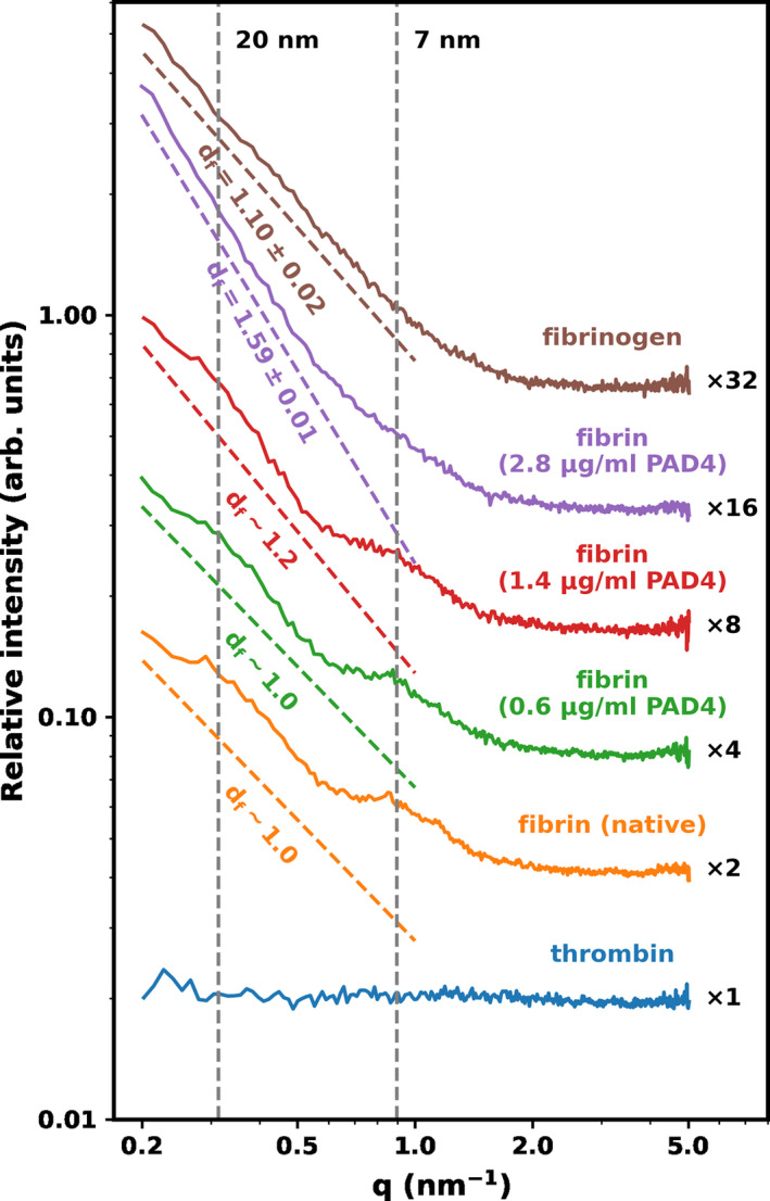
Methods: Fibrinogen was citrullinated with PAD4 and clotted with thrombin. Scanning electron microscopy (SEM) and atomic force microscopy (AFM) were used to measure fiber thickness, fiber height/width ratio, and fiber persistence length in clots containing citFg. Fiber density was measured with laser scanning microscopy (LSM) and permeability measurements were carried out to estimate the porosity of the clots. The intra-fiber structure of fibrin was analyzed with small-angle X-ray scattering (SAXS).

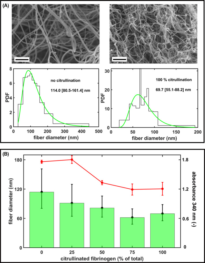
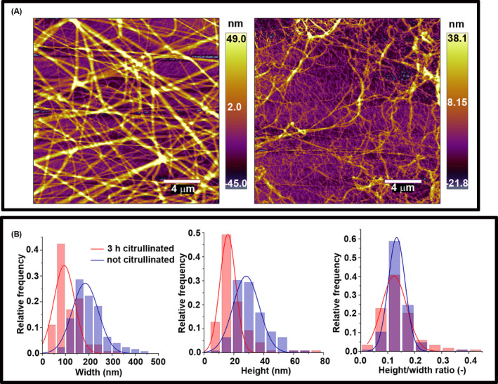
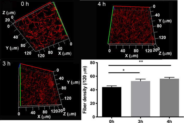
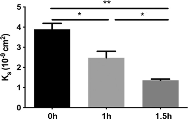
Results: SEM images revealed a decrease in the median fiber diameter that correlated with the fraction of citFg in the clot, while the fiber width/length ratio remained unchanged according to AFM. With SAXS we observed that citrullination resulted in the formation of denser clots in line with increased fiber density shown by LSM. The permeability constant of citrullinated fibrin decreased more than 3-fold indicating significantly decreased porosity. SAXS also showed largely preserved periodicity in the longitudinal assembly of fibrin monomers.

Conclusion: The current observations of thin fibers combined with dense packing and low porosity in the presence of citFg can provide a structural framework for the mechanical fragility and lytic resistance of citrullinated fibrin.

Keywords: atomic force microscopy; citrullination; fibrin; protein-arginine deiminases; small-angle X-ray scattering.


Conflict of interest statement

The authors declare no competing financial interests.

Figures

FIGURE 1
FIGURE 1
Structural effects of fibrinogen citrullination. A, Representative scanning electron microscropy (SEM) images of fibrin clots prepared from 4 μM fibrinogen pre‐treated with 1.15 μg/ml peptidyl arginine deiminase 4 (PAD4) for 0/4 h (clotted with 15 nM thrombin for 2 h at 37°C). Scale bar = 2 μm. The diameter of 300 fibers per image was measured from three SEM images per clot type using the algorithms described in the Materials and Methods section and their empirical (histogram) and fitted theoretical distribution (green line) are shown (PDF, probability density function of the distribution). Numbers indicate the median (bottom‐top quartile) values of fiber diameter. B, Fibrinogen pre‐treated with 1.15 μg/ml PAD4 for 4 h was mixed with non‐citrullinated fibrinogen (containing the PAD4 at the same concentration, but inactivated with 60 μg/ml Cl‐amidine) at the indicated ratios, clotted with thrombin and SEM images were taken as in (A). The bars show the median values, the whiskers span the interquartile range (bottom and quartile values) of the fitted theoretical distributions. All differences are significant at p < .05 level according to Kuiper's test of the distributions. The red symbols represent the maximal absorbance measured at 340 nm in the turbidimetric clotting assay of the indicated fibrinogen preparations (mean ± standard deviation, one‐way analysis of variance p = .03, n = 5).
FIGURE 2
FIGURE 2
Atomic force microscopy studies of citrullinated fibrinogen. A, Fibrinogen at 3 μM citrullinated with 0.25 μg/ml peptidyl arginine deiminase 4 for 0/3 h; left and right, respectively clotted with 10 nM thrombin in a total volume of 20 μl on a mica surface. Scale bar = 4 μm. B, Topographical analysis of the cross‐sectional profile of the fibrin fibers. Fiber height and width were obtained as described in the Materials and Methods section. Further quantitative data calculated from similar images are presented in Tables 1 and 2.
FIGURE 3
FIGURE 3
Fibrin fiber density. Fibrinogen at 3.6 μM (citrullinated with 0.4 μg/ml peptidyl arginine deiminase 4 for the indicated times) was clotted with 16 nM thrombin in the presence of 0.05 μM Alexa Fluor® 546‐conjugated fibrinogen. Images were taken after 30 min incubation at room temperature. Optical z‐stacks (every 1 μm over 20 μm) were combined to construct 3D images. Fiber density was determined by counting the number of fibers crossing 120 μm long sections of 2D‐projected laser scanning microscopy images. Error bars represent standard error of the mean. Each clot was prepared in duplicate, and two density measurements were performed in each (n = 4). *p < .05, **p < .01.
FIGURE 4
FIGURE 4
Fibrin permeability. Permeability of clots prepared from fibrinogen at 8 μM pretreated with 0.6 μg/ml peptidyl arginine deiminase 4 for the indicated times and clotted with 16 nM thrombin. The permeability coefficient (K S) was calculated as described in the Materials and Methods section. K S values and standard error of mean were calculated from at least eight samples originating from three independent experiments. *p < .01, **p < .0001.
FIGURE 5
FIGURE 5
Effect of citrullination on the subfibrillar structure of fibrin. Fibrin clots were formed with fibrinogen pre‐treated with 8 μΜ citrullinated by 0/0.6/1.4/2.8 μg/ml peptidyl arginine deiminase 4 for 1 h and clotted with 10 nM thrombin and examined with small‐angle X‐ray scattering as described in the Materials and Methods section. Fibrinogen and thrombin solutions were used as a reference. The normalized scattering intensity (d∑/d) is plotted as a function of the momentum transfer (q) and empirical curves are fitted to the raw data, as described in the Materials and Methods section. The slope of the dashed color lines indicates the approximated mass fractal dimension of the fibers (d f, best fit of the slope and its uncertainty). For the scattering curves with a diffuse peak at q = .32 lines are fitted to the flanking regions of the peak and approximate d f values are shown. Curves are shifted vertically by the indicated factors for better visualization. The vertical dashed lines indicate the positions of the scattering peaks corresponding to periodicity of ∼7 nm, and 20 nm. The scattering curve of thrombin is also presented to demonstrate the negligibility of the scattering contribution of this component.

References

    1. WHO . World Health Statistics Full report 2021. Accessed April 15, 2022 https://www.who.int/data/gho/publications/world‐health‐statistics
    1. Ariëns RA. Fibrin(ogen) and thrombotic disease. J Thromb Haemost. 2013;11(Suppl 1):294‐305. - PubMed
    1. Longstaff C, Kolev K. Basic mechanisms and regulation of fibrinolysis. J Thromb Haemost. 2015;13(Suppl 1):S98‐S105. - PubMed
    1. Martinez MR, Cuker A, Mills AM, et al. Enhanced lysis and accelerated establishment of viscoelastic properties of fibrin clots are associated with pulmonary embolism. Am J Physiol Lung Cell Mol Physiol. 2014;306:L397‐L404. - PMC - PubMed
    1. Lee JS, Moon T, Kim TH, et al. Deep vein thrombosis in patients with pulmonary embolism: prevalence, clinical significance and outcome. Vasc Specialist Int. 2016;32:166‐174. - PMC - PubMed

Publication types