. 2022 Sep 30;4(1):19-27.
doi: 10.1530/VB-22-0006. Print 2022 Sep 1.

Molecular mechanisms and effects of urocortin II on rat adventitial fibroblast calcification induced by calcified medium

Affiliations

Molecular mechanisms and effects of urocortin II on rat adventitial fibroblast calcification induced by calcified medium

Xusheng Zhang et al. Vasc Biol. .

Abstract

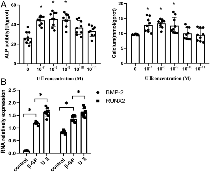
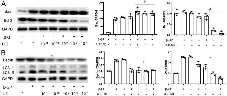
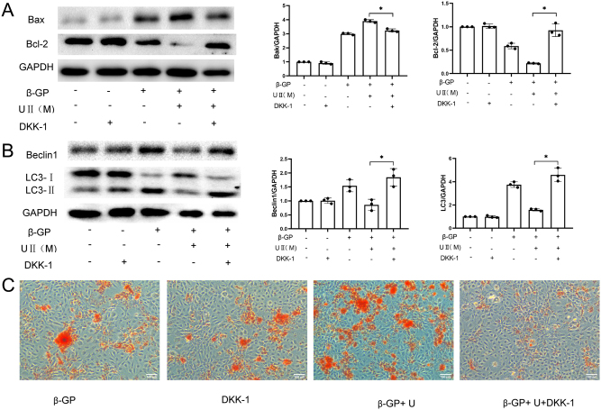
The present study aimed to assess the role of urocortin II (UII) in the process of vascular calcification in vitro by using a calcification model, to detect the changes in the mRNA and protein levels of associated markers in rat adventitial fibroblasts (AFs) during their phenotypic transformation to osteoblast cellsto clarify the main signal transduction pathway of UII responsible for regulating vascular calcification and AF phenotypic transformation of osteoblast cells, and to prove that UII was an endogenous factor promoting vascular calcification, so as to provide an effective experimental basis for the clinical regulation of related diseases caused by vascular calcification. Finally, we successfully constructed the calcified cell model, found that UII was an endogenous substance regulating vascular calcification, regulated the vascular calcification by promoting apoptosis and inhibiting autophagy through up- and downregulated BAX and BCL-2/BECLIN 1 (BECN1) level, and the Wnt/β-catenin signaling pathway was involved.

Keywords: Wnt/β-catenin signaling pathway; rat adventitial fibroblasts; urocortin II; vascular calcification.


Figures

Figure 1
Figure 1
Rat extravascular outer membrane cells induce calcification in vitro. (A) By using the calcification-induced medium, the ALP activity and the intracellular calcium content were increased following the time of culture. (B) In culture, intracellular calcium nodules were clearly observed for 15 days with cissarin red staining (100×). In each group, the cells were seeded into six parallel wells for every single index detection.
Figure 2
Figure 2
UII aggravates the calcification of vascular outer membrane cells in rats. (A) The incubation of the cells with different concentrations of UII demonstrated that high concentrations of UII could increase ALP activity and intracellular calcium content. (B) The expression levels of specific osteogenesis-related genes were detected using the RT-PCR assay, and the data indicated that the levels between all four osteogenesis-related genes were upregulated at 15 days following induction of calcification. UII caused an upregulation in the expression levels of osteogenesis-related genes. In each group, the cells were seeded into six parallel wells for every single index detection.
Figure 3
Figure 3
UII promotes cell calcification by inducing apoptosis and inhibiting autophagy. WB analysis indicated that following 15 days of calcification, the expression levels of the apoptotic protein BAX were increased, whereas those of the antiapoptotic protein BCL-2 were decreased. The expression levels of the autophagy-related proteins BECLIN 1 and LC3 were upregulated. UII caused further upregulation of the expression levels of the apoptotic protein BAX and downregulation of the expression levels of the apoptotic protein BCL-2. The expression levels of the autophagy-related proteins Beclin1 and LC3 were downregulated. The data indicated that UII exacerbated cell calcification by promoting apoptosis and inhibiting autophagy. In each group, the cells were seeded into six parallel wells for every single index detection.
Figure 4
Figure 4
DKK-1, the inhibitor of the Wnt/β-catenin pathway, inhibits the effect of UII. (A and B) WB analysis indicated that treatment of cells with the Wnt/β-catenin inhibitor DKK-1 reversed the effects of UII, causing downregulation of the apoptotic protein BAX, upregulation of the antiapoptotic protein BCL-2, and upregulation of the autophagy-related proteins BECLIN 1 and LC3. (C) Sissarin red staining demonstrated that significant calcium nodules were observed in the intracellular fraction following 15 days of induction with calcified media. The simultaneous addition of the UII culture indicated significantly denser intracellular calcium nodules. The increase in the concentration levels of DKK-1 resulted in significantly smaller orange calcium nodules being present in the cells. In each group, the cells were seeded into six parallel wells for every single index detection.

References

    1. Shanahan CM, Crouthamel MH, Kapustin A, Giachelli CM. Arterial calcification in chronic kidney disease: key roles for calcium and phosphate. Circulation Research 2011109697–711. (10.1161/CIRCRESAHA.110.234914) - DOI - PMC - PubMed
    1. Ho CY, Shanahan CM. Medial arterial calcification: an overlooked player in peripheral arterial disease. Arteriosclerosis, Thrombosis, and Vascular Biology 2016361475–1482. (10.1161/ATVBAHA.116.306717) - DOI - PubMed
    1. Nicoll R, Henein MY. The predictive value of arterial and valvular calcification for mortality and cardiovascular events. International Journal of Cardiology. Heart and Vessels 201431–5. (10.1016/j.ijchv.2014.02.001) - DOI - PMC - PubMed
    1. Johnson RC, Leopold JA, Loscalzo J. Vascular calcification: pathobiological mechanisms and clinical implications. Circulation Research 2006991044–1059. (10.1161/01.RES.0000249379.55535.21) - DOI - PubMed
    1. Yamada S, Giachelli CM. Vascular calcification in CKD-MBD: roles for phosphate, FGF23, and klotho. Bone 201710087–93. (10.1016/j.bone.2016.11.012) - DOI - PMC - PubMed