. 2022 Nov 1;323(5):H917-H933.
doi: 10.1152/ajpheart.00253.2022. Epub 2022 Sep 9.

Role of Axl in target organ inflammation and damage due to hypertensive aortic remodeling

Affiliations

Role of Axl in target organ inflammation and damage due to hypertensive aortic remodeling

Wei Chen et al. Am J Physiol Heart Circ Physiol. .

Abstract

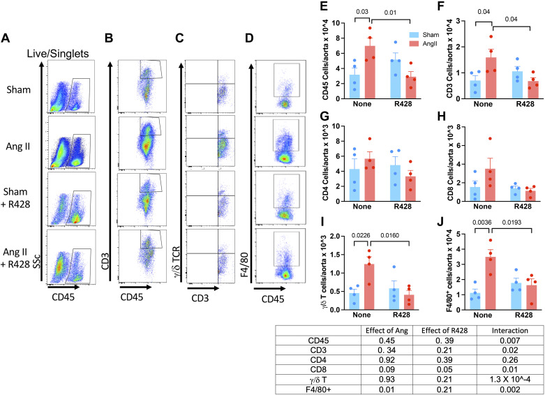
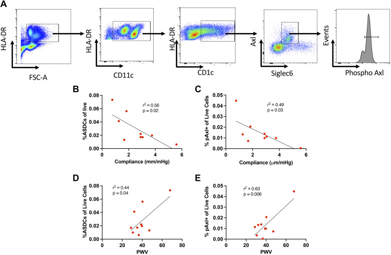
We have shown that excessive endothelial cell stretch causes release of growth arrest-specific 6 (GAS6), which activates the tyrosine kinase receptor Axl on monocytes and promotes immune activation and inflammation. We hypothesized that GAS6/Axl blockade would reduce renal and vascular inflammation and lessen renal dysfunction in the setting of chronic aortic remodeling. We characterized a model of aortic remodeling in mice following a 2-wk infusion of angiotensin II (ANG II). These mice had chronically increased pulse wave velocity, and their aortas demonstrated increased mural collagen. Mechanical testing revealed a marked loss of Windkessel function that persisted for 6 mo following ANG II infusion. Renal function studies showed a reduced ability to excrete a volume load, a progressive increase in albuminuria, and tubular damage as estimated by periodic acid Schiff staining. Treatment with the Axl inhibitor R428 beginning 2 mo after ANG II infusion had a minimal effect on aortic remodeling 2 mo later but reduced the infiltration of T cells, γ/δ T cells, and macrophages into the aorta and kidney and improved renal excretory capacity, reduced albuminuria, and reduced evidence of renal tubular damage. In humans, circulating Axl+/Siglec6+ dendritic cells and phospho-Axl+ cells correlated with pulse wave velocity and aortic compliance measured by transesophageal echo, confirming chronic activation of the GAS6/Axl pathway. We conclude that brief episodes of hypertension induce chronic aortic remodeling, which is associated with persistent low-grade inflammation of the aorta and kidneys and evidence of renal dysfunction. These events are mediated at least in part by GAS6/Axl signaling and are improved with Axl blockade.NEW & NOTEWORTHY In this study, a brief, 2-wk period of hypertension in mice led to progressive aortic remodeling, an increase in pulse wave velocity, and evidence of renal injury, dysfunction, and albuminuria. This end-organ damage was associated with persistent renal and aortic infiltration of CD8+ and γ/δ T cells. We show that this inflammatory response is likely due to GAS6/Axl signaling and can be ameliorated by blocking this pathway. We propose that the altered microvascular mechanical forces caused by increased pulse wave velocity enhance GAS6 release from the endothelium, which in turn activates Axl on myeloid cells, promoting the end-organ damage associated with aortic stiffening.

Keywords: T cells; Windkessel function; aorta; macrophages; vascular remodeling.


Conflict of interest statement

No conflicts of interest, financial or otherwise, are declared by the authors.

Figures

None
Graphical abstract
Figure 1.
Figure 1.
Effects of a prior 2-wk episode of hypertension on aortic morphology. A: blood pressures measured at 1 wk and 2, 4, and 6 mo following a 2-wk infusion of ANG II (n = 5–8 per group). B: examples of hematoxylin and eosin, Picrosirius red, and Masson’s trichrome staining of sections from the perfusion-fixed thoracic aorta from sham and ANG II mice at 2, 4, and 6 mo following ANG II infusion. C: quantifies the adventitial area, obtained from planimetry of Masson’s trichrome images (n = 5–9). D and E: examples of smooth muscle α-actin staining and quantification of medial area from these images (n = 5–9). Table: results of overall comparisons that were performed using aligned rank transformation analysis of variance. Between-group comparisons were made with a Mann–Whitney test followed by a Bonferroni correction. P values reflect this correction.
Figure 2.
Figure 2.
Short- and long-term effects of a 2-wk episode of hypertension on aortic mechanics. Mice underwent a 2-wk infusion of ANG II as in Fig. 1, and aortas were studied as previously described. Comparisons are shown for immediately after and 2, 4, and 6 mo following a 2-wk infusion of either vehicle (sham) or ANG II for aortic loaded internal diameter (A), circumferential stiffness (B), loaded thickness (C), and stored energy (D). Values are n = 5 for each group, and overall comparisons were performed using two-way ANOVA. When significance was indicated, a Sidak’s multiple comparison was performed to detect differences between groups.
Figure 3.
Figure 3.
Transcriptomic changes in aortic gene expression following a 2-wk episode of hypertension. Values are from RNA sequencing from aortas of four mice in each group. A–C: volcano plots of significantly up- and downregulated genes. Gray line delineates false discovery rate of 0.10. D: Venn diagram summarizing the number of genes that were significantly different between sham- and ANG II-infused mice at ×3. E: heat maps of the top 20 up- and downregulated genes at 2 and 4 mo.
Figure 4.
Figure 4.
Persistent effects of hypertension on renal function. A: response to a volume challenge. Mice were injected with normal saline (ip) and immediately placed in a metabolic chamber for 4 h with urine output measured. B: urinary albumin/creatinine levels determined using ELISAs. C and D: representative periodic acid Schiff (PAS) stains for renal tubular injury. E: mean values for PAS scores at 2, 4, and 6 mo. Table: results of the aligned rank transformation ANOVA for these parameters. When between-group significance was indicated, a post hoc Mann–Whitney test followed by a Bonferroni correction was performed (n = 6–8).
Figure 5.
Figure 5.
Effect of Axl blockade on blood pressure and aortic mechanics. Mice underwent a 2-wk infusion of ANG II; then, 2 mo after cessation of this infusion, R428 was added to the drinking water or not for the ensuing 2 mo. A: systolic blood pressure estimated by tail-cuff measures (n = 5). B: examples of Elastica van Gieson’s stains (top) and birefringent images of Picrosirius stains of aortas. C–F: quantification of elastin and collagen areas (n = 5). Values were compared using an unpaired t test. G–J: parameters of aortic mechanics in sham- and ANG II-treated mice with and without R428 treatment (n = 5 for each). K: pulse wave velocity (PWV) measured by Doppler ultrasound in anesthetized mice (n = 2 or 3 for each). Table: results of overall comparisons that were performed using aligned rank transformation analysis of variance. Between-group comparisons were made with a MannWhitney test followed by a Bonferroni correction.
Figure 6.
Figure 6.
Aortic-infiltrating cells 4 mo after a 2-wk episode of hypertension and effect of Axl blockade. Single-cell suspensions from aortic homogenates were subjected to flow cytometry. AD: example flow plots. Mean data ± SE are compared between mice that had received ANG II vs. sham infusions with or without Axl blockade with R428 for total leukocytes (CD45; E), T cells (CD3; F), CD4+ T cells (G), CD8+ T cells (H), γ/δ T cells (I), and F4/80+ cells (J). Table: results of aligned rank transformation and subsequent two-way ANOVA. Data are n = 4 for all.
Figure 7.
Figure 7.
Intracellular staining for IFNγ and IL-17A in subsets of aortic-infiltrating T cells. Mean data ± SE for aortic cells from mice that had received either sham or ANG II infusion with and without treatment with the Axl inhibitor R428 for IFNg in CD4+, CD8+, and γ/δ T cells (A, C, and D) and IL-17A in CD4+ and γ/δ T cells (B and E). Data represent further analysis of samples from Fig. 6 and were analyzed with aligned rank transformed ANOVA and a MannWhitney post hoc test with Bonferroni correction. Data are n = 4 for all.
Figure 8.
Figure 8.
Flow cytometric analysis of renal-infiltrating cells 4 mo after a 2-wk episode of hypertension and effect of Axl blockade. Single-cell suspensions of renal homogenates were stained for the indicated surface markers and then fixed and permeabilized for assessment of intracellular levels of IFNγ and IL-17A. Mean data ± SE are compared between mice that had received ANG II vs. sham infusions with or without Axl blockade with R428 for total leukocytes (CD45; A), T cells (CD3; B), CD4+ T cells (C), CD8+ T cells (D), γ/δ T cells (E), F4/80+ cells (F), IFNg+ γ/δ T cells (G), and IL-17A+ γ/δ T cells (H). Table: results of aligned rank transformation ANOVA. Data are n = 5 or 6 for all.
Figure 9.
Figure 9.
Evidence on myeloid/T-cell interaction in aortas and kidneys of mice 4 mo after ANG II-induced hypertension and effect of R428. A: adjusted forward scatter/side scatter gating. B: presence of clusters of F4/80+ cells with all T cells, CD4+ T cells, CD8+ T cells, and CD4+/CD8+-clustered cells. C: a similar analysis for renal cells. Table: results of aligned rank transformation two-way ANOVA. Data are n = 5 or 6 for all.
Figure 10.
Figure 10.
Prolonged evidence of renal injury and dysfunction following an episode of hypertension and improvement with Axl blockade. A: urinary neutrophil gelatinase-associated lipocalin levels measured by enzyme-linked immunoassays. B: urine excretion of a volume challenge. C and D: example of tubular injury and the tubular injury score as assessed by periodic acid Schiff (PAS) staining. Table: results of overall comparisons that were performed using aligned rank transformation ANOVA. Between-group comparisons were made with a MannWhitney test followed by a Bonferroni correction. P values reflect this correction.
Figure 11.
Figure 11.
Relationship of aortic compliance and pulse wave velocity (PWV) to Axl+/Siglec6+ dendritic cells (ASDCs) and phospho-Axl (pAxl)+ cells. Transesophageal echocardiograms and measures of PWV were obtained in nine patients during cardiac surgery. Simultaneously collected blood samples were analyzed for circulating ASDCs and pAxl+ cells. A: gating strategy for live/singlet cells. B and C: relationships of ASDCs and %pAxl+ live cells to aortic compliance. D and E: relationships of ASDCs and %pAxl+ live cells to PWV.

References

    1. Adji A, O'Rourke MF, Namasivayam M. Arterial stiffness, its assessment, prognostic value, and implications for treatment. Am J Hypertens 24: 5–17, 2011. doi:10.1038/ajh.2010.192. - DOI - PubMed
    1. O'Rourke MF, Hashimoto J. Mechanical factors in arterial aging: a clinical perspective. J Am Coll Cardiol 50: 1–13, 2007. doi:10.1016/j.jacc.2006.12.050. - DOI - PubMed
    1. Safar ME. Arterial aging–hemodynamic changes and therapeutic options. Nat Rev Cardiol 7: 442–449, 2010. doi:10.1038/nrcardio.2010.96. - DOI - PubMed
    1. Mitchell GF, Guo CY, Benjamin EJ, Larson MG, Keyes MJ, Vita JA, Vasan RS, Levy D. Cross-sectional correlates of increased aortic stiffness in the community: the Framingham Heart Study. Circulation 115: 2628–2636, 2007. doi:10.1161/CIRCULATIONAHA.106.667733. - DOI - PubMed
    1. Dietrich T, Schaefer-Graf U, Fleck E, Graf K. Aortic stiffness, impaired fasting glucose, and aging. Hypertension 55: 18–20, 2010. doi:10.1161/HYPERTENSIONAHA.109.135897. - DOI - PubMed

Publication types