Review
. 2022 Nov-Dec;42(7):1925-1939.
doi: 10.1148/rg.220073. Epub 2022 Sep 9.

Interstitial Lung Abnormalities at CT: Subtypes, Clinical Significance, and Associations with Lung Cancer

Affiliations
Review

Interstitial Lung Abnormalities at CT: Subtypes, Clinical Significance, and Associations with Lung Cancer

Akinori Hata et al. Radiographics. 2022 Nov-Dec.

Abstract

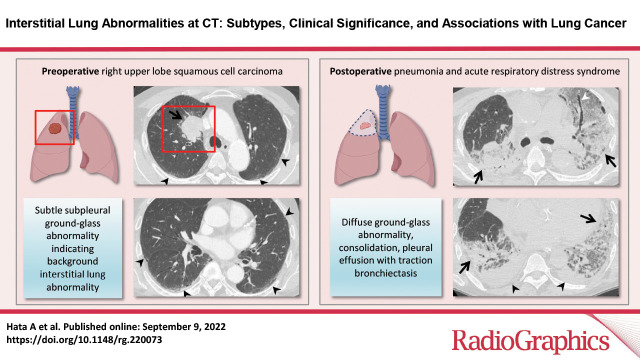
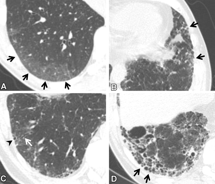
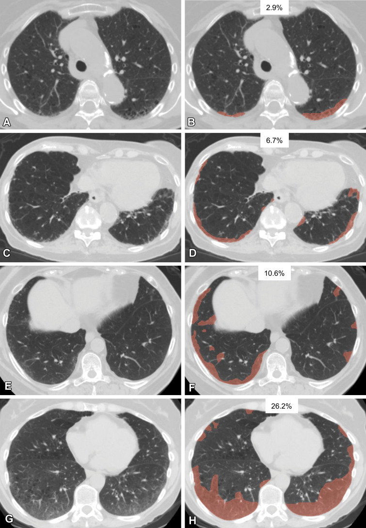
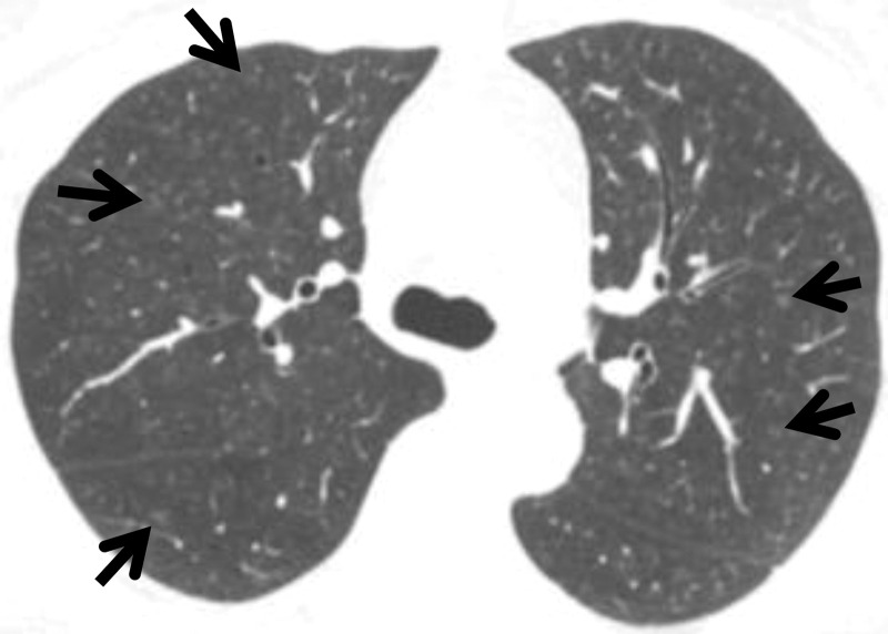
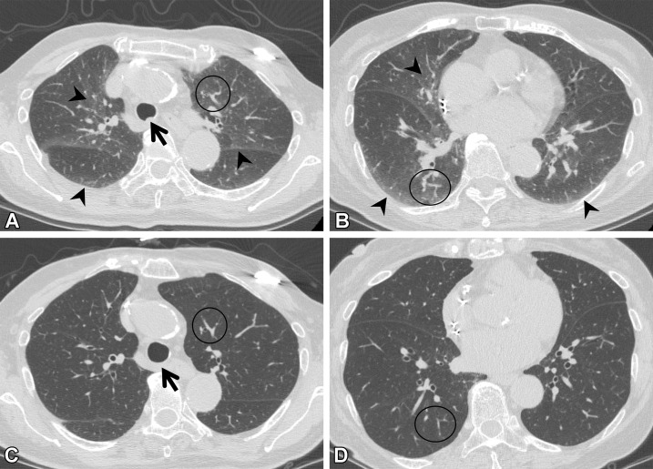
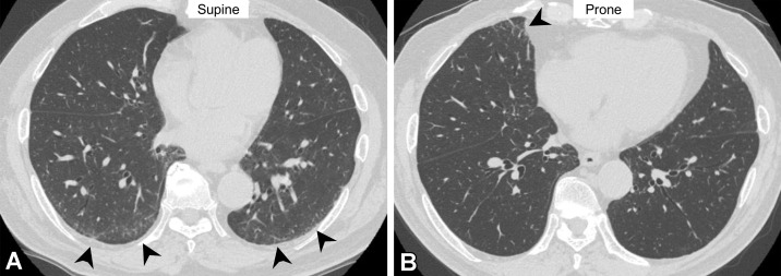
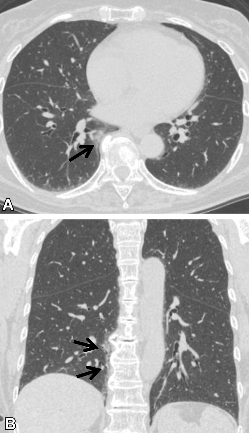
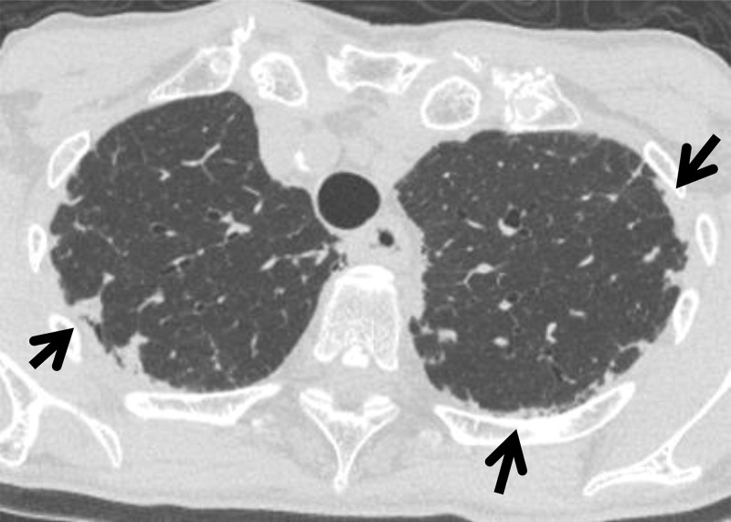
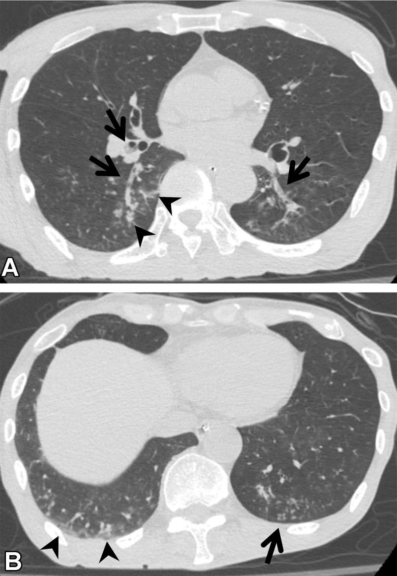
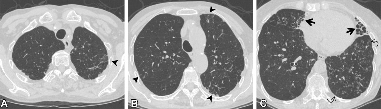
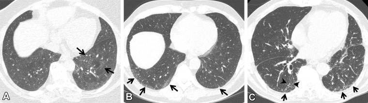
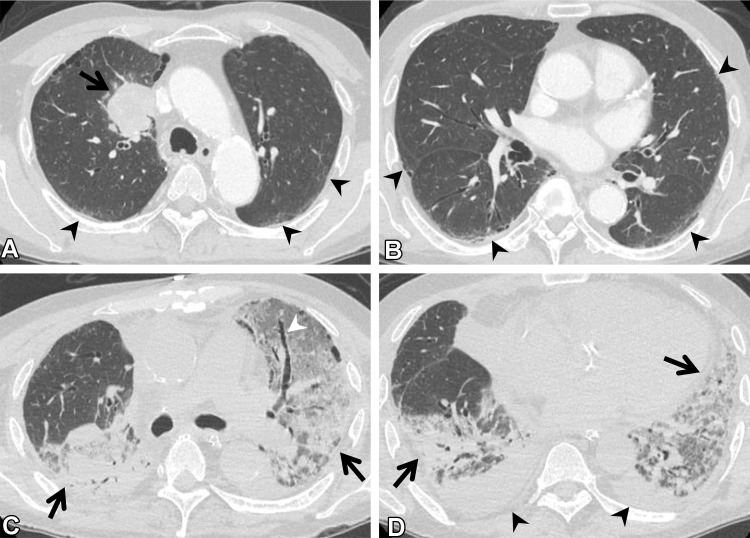
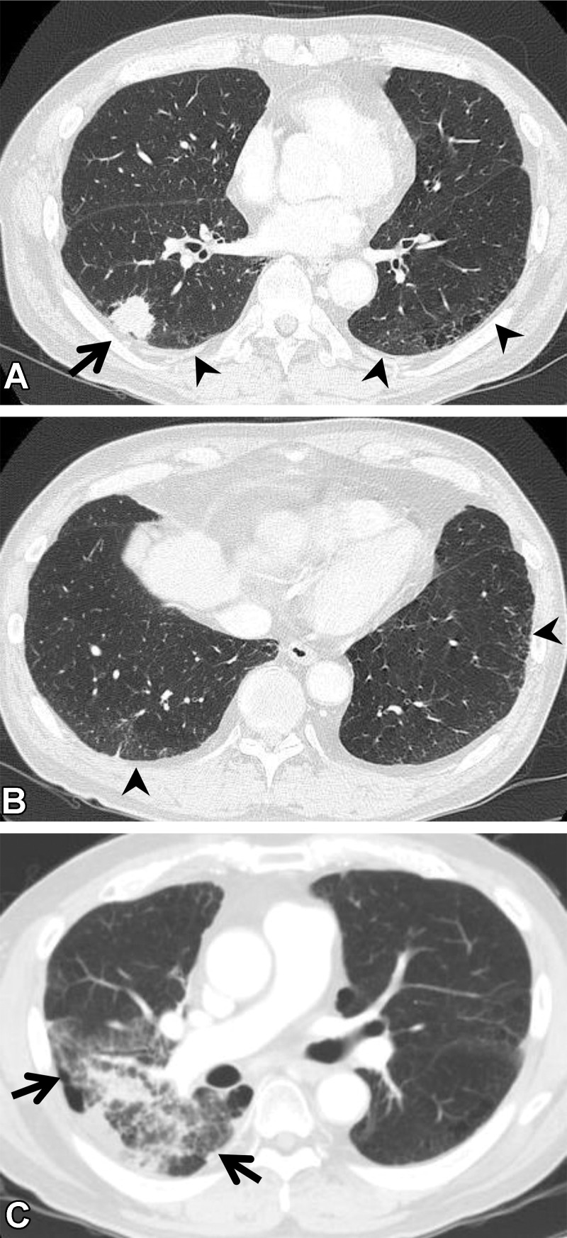
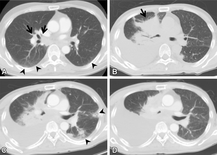
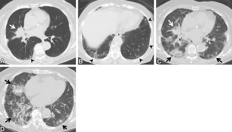
Interstitial lung abnormality (ILA) is defined as an interstitial change detected incidentally on CT images. It is seen in 4%-9% of smokers and 2%-7% of nonsmokers. ILA has a tendency to progress with time and is associated with respiratory symptoms, decreased exercise capability, reduced pulmonary function, and increased mortality. ILAs can be classified into three subcategories: nonsubpleural, subpleural nonfibrotic, and subpleural fibrotic. In cases of ILA, clinically significant interstitial lung disease should be identified and requires clinically driven management by a pulmonologist. Risk factors for the progression of ILA include clinical elements (ie, inhalation exposures, medication use, radiation therapy, thoracic surgery, physiologic findings, and gas exchange findings) and radiologic elements (ie, basal and peripheral predominance and fibrotic findings). It is recommended that individuals with one or more clinical or radiologic risk factors for progression of ILA be actively monitored with pulmonary function testing and CT. To avoid overcalling ILA at CT, radiologists must recognize the imaging pitfalls, including centrilobular nodularity, dependent abnormality, suboptimal inspiration, osteophyte-related lesions, apical cap and pleuroparenchymal fibroelastosis-like lesions, aspiration, and infection. There is a close association between ILA and lung cancer, and many studies have reported an increased incidence of lung cancer, worse prognoses, and/or increased pulmonary complications in relation to cancer treatment in patients with ILA. ILA is considered to be an important comorbidity in patients with lung cancer. Accordingly, all radiologists involved with body CT must have sound knowledge of ILAs owing to the high prevalence and potential clinical significance of these anomalies. An overview of ILAs, including a literature review of the associations between ILAs and lung cancer, is presented. ©RSNA, 2022.


Conflict of interest statement

Disclosures of conflicts of interest.—: M.N. Institutional research grants from Canon Medical Systems, Daiichi Sankyo, and AstraZeneca; consulting fees from Daiichi Sankyo and AstraZeneca. G.M.H. Consulting fees from Boehringer-Ingelheim, Chugai Pharmaceuticals, and Gerson Lehrman Group; participation on Data Safety Monitoring Board or Advisory Board of Biogen. H.H. Institutional research grants from Canon Medical Systems and Konica-Minolta; personal consulting fees from Mitsubishi Chemical and Canon Medical Systems.

Figures

None
Created with BioRender.com
CT findings that constitute ILAs. Axial images show ground-glass
abnormality (arrows in A), reticulation (arrows in B), traction
bronchiectasis (arrow in C), nonemphysematous cyst (arrowhead in C), and
honeycombing (arrows in D).
Figure 1.
CT findings that constitute ILAs. Axial images show ground-glass abnormality (arrows in A), reticulation (arrows in B), traction bronchiectasis (arrow in C), nonemphysematous cyst (arrowhead in C), and honeycombing (arrows in D).
Various extents of lung abnormality in four patients. Original axial
CT images (A, C, E, G) and the same CT images with a segmented overlay of
ILA (B, D, F, and H) are shown. (A, B) Scans in a 76-year-old woman show
subtle subpleural ground-glass abnormality that accounts for 2.9% of the
lung area, indicating insignificant abnormality. (C, D) Scans in an
84-year-old woman show ground-glass and reticular abnormalities that account
for 6.7% of the lung area, indicating significant abnormality. (E, F) Scans
in a 73-year-old woman show faint ground-glass abnormality that accounts for
10.6% of the lung area, indicating significant abnormality. (G, H) Scans in
a 60-year-old woman show widespread ground-glass abnormality that affects
26.2% of the lung area, indicating significant abnormality. The
segmentations shown were performed manually by using 3D Slicer software
(Brigham and Women’s Hospital, Harvard Medical School, Boston,
MA).
Figure 2.
Various extents of lung abnormality in four patients. Original axial CT images (A, C, E, G) and the same CT images with a segmented overlay of ILA (B, D, F, and H) are shown. (A, B) Scans in a 76-year-old woman show subtle subpleural ground-glass abnormality that accounts for 2.9% of the lung area, indicating insignificant abnormality. (C, D) Scans in an 84-year-old woman show ground-glass and reticular abnormalities that account for 6.7% of the lung area, indicating significant abnormality. (E, F) Scans in a 73-year-old woman show faint ground-glass abnormality that accounts for 10.6% of the lung area, indicating significant abnormality. (G, H) Scans in a 60-year-old woman show widespread ground-glass abnormality that affects 26.2% of the lung area, indicating significant abnormality. The segmentations shown were performed manually by using 3D Slicer software (Brigham and Women’s Hospital, Harvard Medical School, Boston, MA).
Diagram shows the relationship between ILA and ILD.
Figure 3.
Diagram shows the relationship between ILA and ILD.
Centrilobular nodularity in a 50-year-old woman who currently smoked
and had a history of 35 pack-years without respiratory symptoms. Axial CT
image shows small nodules (arrows) with centrilobular distribution in the
bilateral lungs.
Figure 4.
Centrilobular nodularity in a 50-year-old woman who currently smoked and had a history of 35 pack-years without respiratory symptoms. Axial CT image shows small nodules (arrows) with centrilobular distribution in the bilateral lungs.
Suboptimal inspiration in an 87-year-old woman who underwent CT for
evaluation of a lung nodule. (A, B) Axial CT images show ground-glass
abnormalities (arrowheads) in subpleural and central areas of the lung zone.
Anterior bulging of the posterior membranous portion of the trachea (arrow
in A) and tortuosity of the vessels (circle) suggest suboptimal inspiration.
(C, D) Follow-up axial CT images show that the ground-glass abnormality has
disappeared, and the normal round shape of the trachea is seen (arrow in C).
Tortuosity of the vessels (circle) is no longer seen, and the lung volume,
as compared with that in A and B, has improved.
Figure 5.
Suboptimal inspiration in an 87-year-old woman who underwent CT for evaluation of a lung nodule. (A, B) Axial CT images show ground-glass abnormalities (arrowheads) in subpleural and central areas of the lung zone. Anterior bulging of the posterior membranous portion of the trachea (arrow in A) and tortuosity of the vessels (circle) suggest suboptimal inspiration. (C, D) Follow-up axial CT images show that the ground-glass abnormality has disappeared, and the normal round shape of the trachea is seen (arrow in C). Tortuosity of the vessels (circle) is no longer seen, and the lung volume, as compared with that in A and B, has improved.
Dependent abnormality in a 72-year-old man with rheumatoid arthritis.
(A) Axial CT image obtained with the patient supine shows ground-glass
abnormality (arrowheads) in the subpleural lung area. (B) On the axial CT
image obtained with the patient prone, the ground-glass abnormality has
disappeared in the subpleural area but is seen in the middle lobe of the
right lung (arrowhead). These findings are considered to indicate transient
lung atelectasis.
Figure 6.
Dependent abnormality in a 72-year-old man with rheumatoid arthritis. (A) Axial CT image obtained with the patient supine shows ground-glass abnormality (arrowheads) in the subpleural lung area. (B) On the axial CT image obtained with the patient prone, the ground-glass abnormality has disappeared in the subpleural area but is seen in the middle lobe of the right lung (arrowhead). These findings are considered to indicate transient lung atelectasis.
Osteophyte-related lesion in a 72-year-old woman with no respiratory
symptoms. (A) Axial CT image shows ground-glass abnormality adjacent to an
osteophyte (arrow). (B) Coronal CT image shows the craniocaudal alignment
(arrows) of the abnormality.
Figure 7.
Osteophyte-related lesion in a 72-year-old woman with no respiratory symptoms. (A) Axial CT image shows ground-glass abnormality adjacent to an osteophyte (arrow). (B) Coronal CT image shows the craniocaudal alignment (arrows) of the abnormality.
PPFE-like lesion in a 73-year-old woman. Axial CT image shows
subpleural soft-tissue attenuation with irregular borders (arrows) in the
upper lungs. There was no interval change in this abnormality at
follow-up.
Figure 8.
PPFE-like lesion in a 73-year-old woman. Axial CT image shows subpleural soft-tissue attenuation with irregular borders (arrows) in the upper lungs. There was no interval change in this abnormality at follow-up.
Aspiration in a 64-year-old man with corticobasal degeneration. (A)
Axial CT image shows central airway plugging (arrows) and centrilobular
nodularity (arrowheads) bilaterally in the lower lung lobes. (B) Axial CT
image at the level of the lung base shows ground-glass abnormality
(arrowheads) in the subpleural area and peripheral centrilobular nodules
(arrow).
Figure 9.
Aspiration in a 64-year-old man with corticobasal degeneration. (A) Axial CT image shows central airway plugging (arrows) and centrilobular nodularity (arrowheads) bilaterally in the lower lung lobes. (B) Axial CT image at the level of the lung base shows ground-glass abnormality (arrowheads) in the subpleural area and peripheral centrilobular nodules (arrow).
Mycobacterium intracellulare infection (non-ILA) in a 72-year-old
woman. (A, B) Axial CT images show linear opacity and ground-glass
abnormality (arrowheads) in the subpleural area of the upper lung lobes. (C)
Axial CT image at a lower level shows bronchiectasis and reticulation
(straight arrows) in the middle lobe and lingula and centrilobular
nodularity (curved arrows) in the left lower lobe. These findings are
typical of nontuberculous mycobacterial pulmonary disease, suggesting that
the abnormalities at the upper level are related to infection or
postinfection change. The findings in this patient were not considered to
indicate ILA.
Figure 10.
Mycobacterium intracellulare infection (non-ILA) in a 72-year-old woman. (A, B) Axial CT images show linear opacity and ground-glass abnormality (arrowheads) in the subpleural area of the upper lung lobes. (C) Axial CT image at a lower level shows bronchiectasis and reticulation (straight arrows) in the middle lobe and lingula and centrilobular nodularity (curved arrows) in the left lower lobe. These findings are typical of nontuberculous mycobacterial pulmonary disease, suggesting that the abnormalities at the upper level are related to infection or postinfection change. The findings in this patient were not considered to indicate ILA.
Subcategories of ILA seen on axial CT images. (A) Nonsubpleural ILA:
faint ground-glass abnormality (arrows) in the central and subpleural areas
of the lungs but not predominant in the subpleural area. (B) Subpleural
nonfibrotic ILA: ground-glass abnormality (arrows) without fibrosis such as
bronchiectasis and architectural distortion. (C) Subpleural fibrotic ILA:
ground-glass abnormality and linear opacities (arrows), with traction
bronchiectasis (arrowheads).
Figure 11.
Subcategories of ILA seen on axial CT images. (A) Nonsubpleural ILA: faint ground-glass abnormality (arrows) in the central and subpleural areas of the lungs but not predominant in the subpleural area. (B) Subpleural nonfibrotic ILA: ground-glass abnormality (arrows) without fibrosis such as bronchiectasis and architectural distortion. (C) Subpleural fibrotic ILA: ground-glass abnormality and linear opacities (arrows), with traction bronchiectasis (arrowheads).
Postoperative complications in a 66-year-old man with squamous cell
cancer. (A, B) Axial preoperative CT images show a mass (arrow in A) in the
right upper lobe of the lung. Subtle ground-glass abnormality (arrowheads),
indicating ILA, is seen in the subpleural area. (C, D) Axial postoperative
CT images show diffuse ground-glass abnormality and consolidation (arrows)
with traction bronchiectasis (arrowhead in C). Pleural effusion (arrowheads
in D) also is seen. The patient was clinically diagnosed to have pneumonia
and acute respiratory distress syndrome.
Figure 12.
Postoperative complications in a 66-year-old man with squamous cell cancer. (A, B) Axial preoperative CT images show a mass (arrow in A) in the right upper lobe of the lung. Subtle ground-glass abnormality (arrowheads), indicating ILA, is seen in the subpleural area. (C, D) Axial postoperative CT images show diffuse ground-glass abnormality and consolidation (arrows) with traction bronchiectasis (arrowhead in C). Pleural effusion (arrowheads in D) also is seen. The patient was clinically diagnosed to have pneumonia and acute respiratory distress syndrome.
RP in a 64-year-old man with squamous cell cancer. (A, B) Axial
pretreatment CT images show a nodule (arrow in A) in the right upper lobe
and ground-glass abnormality (arrowheads) in the subpleural area, suggesting
ILA. (C) Axial CT image after radiation therapy shows ground-glass
abnormality and consolidation (arrows) in the right lung, beyond the
irradiated field. The patient required steroid therapy for RP.
Figure 13.
RP in a 64-year-old man with squamous cell cancer. (A, B) Axial pretreatment CT images show a nodule (arrow in A) in the right upper lobe and ground-glass abnormality (arrowheads) in the subpleural area, suggesting ILA. (C) Axial CT image after radiation therapy shows ground-glass abnormality and consolidation (arrows) in the right lung, beyond the irradiated field. The patient required steroid therapy for RP.
ICI-related pneumonitis in a 62-year-old man with squamous cell
cancer. The patient was treated with nivolumab as the sixth line of therapy.
(A) Axial pretreatment CT image shows subtle ground-glass abnormality
(arrowheads) in the subpleural area, indicating ILA. Right hilar
lymphadenopathy (arrows) also is apparent. (B) Axial CT image 10 months
later shows a mass (arrow) in the right lung and pleural effusion,
suggesting tumor exacerbation. (C) Axial CT image 20 days after the patient
started nivolumab therapy shows a newly emergent patchy ground-glass
abnormality and consolidation (arrowheads), representing ICI-related
pneumonitis with an organizing pneumonia pattern; nivolumab was
discontinued. (D) Axial CT image 3 months after the discontinuation of
nivolumab shows that the patchy opacities have disappeared.
Figure 14.
ICI-related pneumonitis in a 62-year-old man with squamous cell cancer. The patient was treated with nivolumab as the sixth line of therapy. (A) Axial pretreatment CT image shows subtle ground-glass abnormality (arrowheads) in the subpleural area, indicating ILA. Right hilar lymphadenopathy (arrows) also is apparent. (B) Axial CT image 10 months later shows a mass (arrow) in the right lung and pleural effusion, suggesting tumor exacerbation. (C) Axial CT image 20 days after the patient started nivolumab therapy shows a newly emergent patchy ground-glass abnormality and consolidation (arrowheads), representing ICI-related pneumonitis with an organizing pneumonia pattern; nivolumab was discontinued. (D) Axial CT image 3 months after the discontinuation of nivolumab shows that the patchy opacities have disappeared.
Complications of targeted drug therapy in an 85-year-old woman with
adenocarcinoma who was treated with osimertinib. (A, B) Axial pretreatment
CT images show a mass (arrow in A) in the right middle lobe. The
ground-glass abnormality and reticulation (arrowheads) seen in the
subpleural area suggest ILA. The patient’s Krebs von den Lungen 6 and
surfactant protein D levels were normal. (C, D) Axial CT images at 5 months
of treatment show the mass (white arrow in C), and diffuse ground-glass
abnormality and consolidation (black arrows), which indicate pneumonitis.
The patient was short of breath and was treated with steroids.
Figure 15.
Complications of targeted drug therapy in an 85-year-old woman with adenocarcinoma who was treated with osimertinib. (A, B) Axial pretreatment CT images show a mass (arrow in A) in the right middle lobe. The ground-glass abnormality and reticulation (arrowheads) seen in the subpleural area suggest ILA. The patient’s Krebs von den Lungen 6 and surfactant protein D levels were normal. (C, D) Axial CT images at 5 months of treatment show the mass (white arrow in C), and diffuse ground-glass abnormality and consolidation (black arrows), which indicate pneumonitis. The patient was short of breath and was treated with steroids.

References

    1. Hatabu H , Hunninghake GM , Lynch DA . Interstitial lung abnormality: recognition and perspectives . Radiology 2019. ; 291 ( 1 ): 1 – 3 . - PubMed
    1. Hatabu H , Hunninghake GM , Richeldi L , et al . Interstitial lung abnormalities detected incidentally on CT: a position paper from the Fleischner Society . Lancet Respir Med 2020. ; 8 ( 7 ): 726 – 737 . - PMC - PubMed
    1. Hata A , Schiebler ML , Lynch DA , Hatabu H . Interstitial Lung Abnormalities: State of the Art . Radiology 2021. ; 301 ( 1 ): 19 – 34 . - PMC - PubMed
    1. Washko GR , Lynch DA , Matsuoka S , et al . Identification of early interstitial lung disease in smokers from the COPDGene Study . Acad Radiol 2010. ; 17 ( 1 ): 48 – 53 . - PMC - PubMed
    1. Washko GR , Hunninghake GM , Fernandez IE , et al . Lung volumes and emphysema in smokers with interstitial lung abnormalities . N Engl J Med 2011. ; 364 ( 10 ): 897 – 906 . - PMC - PubMed