. 2023 Jan 27;32(4):595-607.
doi: 10.1093/hmg/ddac227.

Multidisciplinary team directed analysis of whole genome sequencing reveals pathogenic non-coding variants in molecularly undiagnosed inherited retinal dystrophies

Affiliations

Multidisciplinary team directed analysis of whole genome sequencing reveals pathogenic non-coding variants in molecularly undiagnosed inherited retinal dystrophies

Malena Daich Varela et al. Hum Mol Genet. .

Abstract

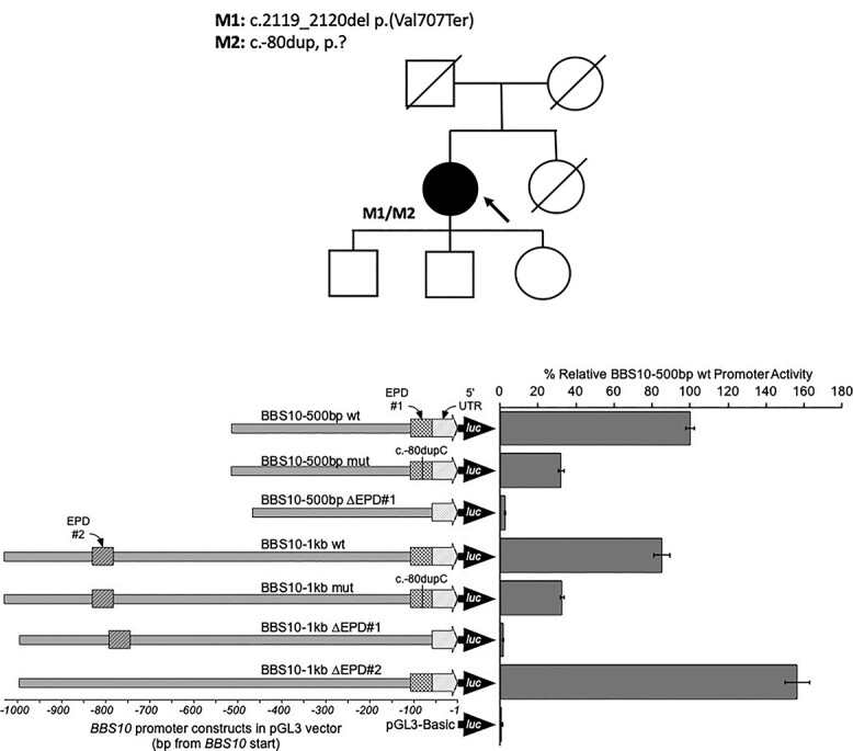
The purpose of this paper is to identify likely pathogenic non-coding variants in inherited retinal dystrophy (IRD) genes, using genome sequencing (GS). Patients with IRD were recruited to the study and underwent comprehensive ophthalmological evaluation and GS. The results of GS were investigated through virtual gene panel analysis, and plausible pathogenic variants and clinical phenotype evaluated by the multidisciplinary team (MDT) discussion. For unsolved patients in whom a specific gene was suspected to harbor a missed pathogenic variant, targeted re-analysis of non-coding regions was performed on GS data. Candidate variants were functionally tested by messenger RNA analysis, minigene or luciferase reporter assays. Previously unreported, likely pathogenic, non-coding variants in 7 genes (PRPF31, NDP, IFT140, CRB1, USH2A, BBS10 and GUCY2D), were identified in 11 patients. These were shown to lead to mis-splicing (PRPF31, IFT140, CRB1 and USH2A) or altered transcription levels (BBS10 and GUCY2D). MDT-led, phenotype-driven, non-coding variant re-analysis of GS is effective in identifying the missing causative alleles.


Figures

Figure 1
Figure 1
Retinal imaging from individuals with IRD within the analyzed cohort. (A) Ultrawide-field (UWF) colour fundus image showing pigmented bone spicules-like lesions and vessel thinning in patient 1, with PRPF31-associated RCD. Fundus autofluorescence (FAF) imaging is positive for a macular hyperautofluorescent ring, characteristic of rod-cone dystrophies (RCD), and generalized decreased hypoAF. Macular optical coherence tomography (OCT) shows a subfoveal island of outer layers. (B) UWF colour and FAF images from patient 4, a child with Norrie disease and bilateral retinal folds. Prophylactic bilateral pan-retinal photocoagulation spots are also visible, marked with red arrows. Macular OCT shows a retinal fold and poor retinal architecture. (C) UWF fundus and OCT imaging from patient 6, who has CRB1-early onset severe retinal dystrophy. Retinal images are positive for nummular, dense, deep pigment deposition, preserved autofluorescence adjacent to few peripheral retinal arterioles and a poorly laminated retina. (D) UWF colour and FAF imaging of patient 7, showing typical RCD features associated with USH2A retinopathy. We can see dense pigment deposits in the mid-periphery, a perifoveal hyperautofluorescent ring demarcating the area of functioning retina, and decreased peripheral AF. Macular OCT shows a subfoveal island of outer segments and cystoid macular oedema. (E) UWF colour, FAF and OCT imaging from patient 8, with BBS10-associated retinopathy. Retinal appearance shows a CRD pattern, with demarcated posterior pole and peripheral areas of retinal pigment epithelium atrophy. Macular OCT appears well correlated, with generalized loss of the outer layers. (F) UWF colour, FAF and OCT images of patient 9, diagnosed with GUCY2D-related EOSRD. Of note is the featureless fundus, mild vessel narrowing, macular atrophy, and disrupted ellipsoid zone centrally.
Figure 2
Figure 2
Analysis of mis-splicing owing to PRPF31 c.1374 + 569C > G. Pedigree of patient 1 showing a dominant family history with incomplete penetrance. Reverse transcription polymerase chain reaction (RT-PCR) and direct sequencing of patient 1 PRPF31 transcript (exon 8–14) derived from PAXgene RNA. Agarose gel analysis showing two distinct bands from RT-PCR amplification in patient sample. L: ladder. Direct sequencing of amplicons shows inclusion of 88 bp pseudoexon leading to a frameshift and premature termination.
Figure 3
Figure 3
Analysis of mis-splicing owing to IFT140 c.2577 + 4_2577 + 5del. Pedigree of patient 3. Nested reverse transcription polymerase chain reaction (RT-PCR) showing multiple fragments amplified from patient 3 cDNA derived from PAXgene RNA sample. Bands [lane 3 (patient) fragment 1 (wild-type, WT), 2 and 3, L: ladder] were purified and sequenced by direct Sanger sequencing. Fragments 2 and 3 showed partial skipping of exon 20 and complete skipping of exon 20, respectively, with clustal alignment of the fragments shown.
Figure 4
Figure 4
Minigene analysis of CRB1 c.3879-1203C > G. Pedigrees of patients 5 and 6. HEK293 cells transfected with CRB1-wt or CRB1-1203C > G containing minigene constructs. The PCR analysis shows splicing in of a 156-bp cryptic exon included in the CRB1-1203C > G transfected cells not present in the wild-type cells.
Figure 5
Figure 5
Cryptic exon inclusion consequent upon USH2A c.4885 + 375A > G. Prediction of pseudoexon inclusion owing to a strengthened deep intronic splice donor site (USH2A is on the reverse strand). Direct Sanger sequencing of PCR amplicons showed an alternate splice product comprising the inclusion of the predicted pseudoexon (130 bp), frameshift and premature stop codon (p.Gly1629ValfsTer52).
Figure 6
Figure 6
BBS10 promoter constructs and activity assayed by firefly luciferase expression in HEK293 cells. The various BBS10 promoter constructs are represented showing the relative positions of the c.-80dupC variant, 5′ UTR, and the EPD#1 and EPD#2 elements. The firefly luciferase reporter (luc) is also indicated. Expression levels are depicted relative to the wild-type BBS10 (500 bp) promoter with 95% confidence intervals indicated. BBS10 (500 bp) promoter variants are indicated by the fine crosshatching, while BBS10 (1 kb) promoter variants are indicated by bold crosshatching. Deletion of the EPD#1 promoter region resulted in complete loss of expression, whereas deletion of EPD#2 resulted in increased transcription (>1.5-fold). The mutant constructs showed an ~70% reduction in expression levels.

References

    1. Pontikos, N., Arno, G., Jurkute, N., Schiff, E., Ba-Abbad, R., Malka, S., Gimenez, A., Georgiou, M., Wright, G., Armengo, M.et al. (2020) Genetic basis of inherited retinal disease in a molecularly characterized cohort of more than 3000 families from the United Kingdom. Ophthalmology, 127, 1384–1394. - PMC - PubMed
    1. Hanany, M., Rivolta, C. and Sharon, D. (2020) Worldwide carrier frequency and genetic prevalence of autosomal recessive inherited retinal diseases. Proc. Natl. Acad. Sci. USA, 117, 2710–2716. - PMC - PubMed
    1. Galvin, O., Chi, G., Brady, L., Hippert, C., Del Valle, R.M., Daly, A. and Michaelides, M. (2020) The impact of inherited retinal diseases in the Republic of Ireland (ROI) and the United Kingdom (UK) from a cost-of-illness perspective. Clin. Ophthalmol., 14, 707–719. - PMC - PubMed
    1. Tsui, I., Song, B.J., Lin, C.S. and Tsang, S.H. (2018) A practical approach to retinal dystrophies. Adv. Exp. Med. Biol., 1085, 245–259. - PubMed
    1. Daich Varela, M., Guimaraes, T., Georgiou, M. and Michaelides, M.. Leber congenital amaurosis/early-onset severe retinal dystrophy: current management and clinical trials. Br. J. Ophthalmol.2022 Apr;106(4), 445–451. - PMC - PubMed

Publication types