. 2022 Sep 15;185(19):3603-3616.e13.
doi: 10.1016/j.cell.2022.08.010. Epub 2022 Aug 29.

Deep mutational scanning identifies SARS-CoV-2 Nucleocapsid escape mutations of currently available rapid antigen tests

Affiliations

Deep mutational scanning identifies SARS-CoV-2 Nucleocapsid escape mutations of currently available rapid antigen tests

Filipp Frank et al. Cell. .

Abstract

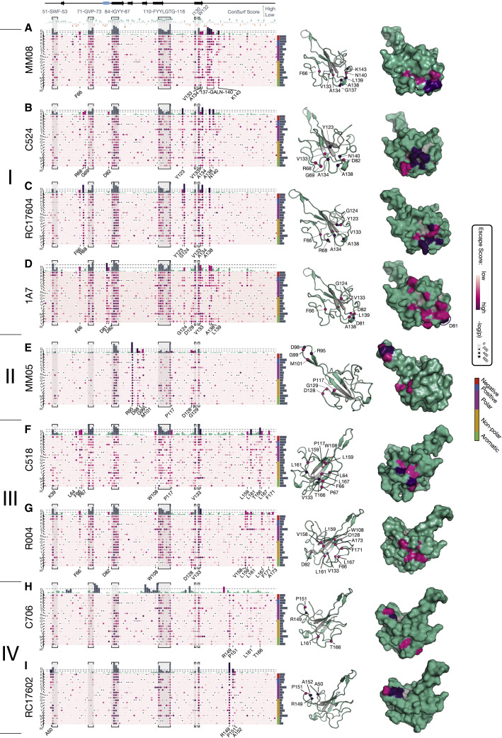
The effects of mutations in continuously emerging variants of SARS-CoV-2 are a major concern for the performance of rapid antigen tests. To evaluate the impact of mutations on 17 antibodies used in 11 commercially available antigen tests with emergency use authorization, we measured antibody binding for all possible Nucleocapsid point mutations using a mammalian surface-display platform and deep mutational scanning. The results provide a complete map of the antibodies' epitopes and their susceptibility to mutational escape. Our data predict no vulnerabilities for detection of mutations found in variants of concern. We confirm this using the commercial tests and sequence-confirmed COVID-19 patient samples. The antibody escape mutational profiles generated here serve as a valuable resource for predicting the performance of rapid antigen tests against past, current, as well as any possible future variants of SARS-CoV-2, establishing the direct clinical and public health utility of our system.

Keywords: SARS-CoV-2 Nucleocapsid protein; antibodies; deep mutational scanning; epitope mapping; mammalian surface-display; rapid diagnostic tests; variants.


Conflict of interest statement

Declaration of interests The authors declare no competing interests.

Figures

None
Graphical abstract
Figure 1
Figure 1
SARS-CoV-2 Nucleocapsid mammalian surface-display platform (A) SARS-CoV-2 Nucleocapsid sequence conservation and disorder prediction (VSL-2). Conservation scores were calculated using ConSurf and 77 coronavirus Nucleocapsid protein sequences. (B) Construct design for mammalian surface-display. A signal peptide and Myc-tag were introduced at the N terminus and a transmembrane helix at the C terminus of the Nucleocapsid protein. The construct was cloned into a lentiviral expression plasmid containing a GFP marker expressed from the same mRNA via an internal ribosomal entry site (IRES). (C) Schematic for detection of surface-displayed Nucleocapsid protein. (D) Flow cytometry analysis of HEK293 cells stably expressing surface-displayed Nucleocapsid. The majority of cells are GFP+ and Myc+ (>90%). GFP+Myc+-gated cells were analyzed for anti-N antibody binding signal (via phycoerythrin [PE]-labeled secondary antibody). Titration experiments for all antibodies used in this study are shown with the normalized median fluorescence intensity (MFI) signal for PE. (E) Validation of dissociation constants determined by mammalian display with dissociation constants from BLI with recombinant protein. Experiments were performed at least twice with similar results for each titration. See also Figure S1.
Figure S1
Figure S1
Gating scheme for antibody titrations using HEK293 cells stably expressing surface-displayed SARS-CoV-2 Nucleocapsid protein, related to Figure 1
Figure S2
Figure S2
Site-saturation library generation and statistics, related to Figure 2 (A) PacBio long-read sequencing statistics showing the length distribution of Nucleocapsid coding sequences, the number of mutations present in sequences with the correct length, and the number of unique barcodes per mutation in reads with the correct length and single mutations. The majority of reads have the correct length of 1,254 nucleotides. Within the sequences with correct length the majority of reads contain a single mutation. Reads with more than one mutation were ignored in all further analyses. (B) Read counts from PacBio sequencing experiments as a tiled heat map. Black points correspond to the wild-type sequence, and pink tiles are mutation without data. (C) Coverage of the Nucleocapsid point mutational space. Shown are the PacBio read depths for all mutations. Missing mutations are shown in pink: residues 251 and 252 were missing from the input library. (D) The lentiviral plasmid libraries were packaged into lentivirus libraries, and HEK293 cells were transduced at an MOI of approximately 0.15 so that 15% of cells were GFP-positive. At least 5 million cells were collected for each replicate library to maintain library complexity. (E) GFP-positive libraries were grown up and then subjected to a second selection step sorting for Myc-positive cells. Approximately 85.9% of cells (shown is library 1) were GFP-positive and 49.2% of cells were GFP-positive and Myc-positive. GFP-positive, but Myc-negative cells made up 36.7% of all cells. (F) Sample gating scheme for DMS screening experiments. (G) Sample data showing escape score transformations. Normalized escape scores resembled a truncated normal distribution. Data were then arcsine square root transformed to generate a symmetrical distribution, followed by Z-normalization to generate data with consistent means and standard deviations between antibody data.
Figure S3
Figure S3
Validation of deep mutational scanning data using individual mutations, related to Figure 3 Shown is a representative gating scheme for antibody titrations using HEK293 cells transfected with constructs for surface-displayed expression of SARS-CoV-2 Nucleocapsid protein mutations.
Figure S4
Figure S4
Full escape mutation profiles of antibodies binding to the dimerization domain, related to Figure 4 The order of amino acids is the same as in the main text figures (from top to bottom: D, E, K, R, H, C, S, T, N, Q, G, A, V, L, I, M, P, F, Y, and W). A subset of antibodies had secondary epitopes at the N-terminal end of the protein (Nab3, Ab166, and mAb 1C1).
Figure 2
Figure 2
Deep mutational scanning approach for determining escape mutations of N-specific antibodies (A) Schematic outlining the deep mutational scanning approach. 15 nucleotide barcodes were added to a site-saturation library containing all point mutations in the Nucleocapsid protein sequence, and the resulting constructs were cloned into a lentiviral expression plasmid (pLVX-IRES-ZsGreen). PacBio long-read sequencing was employed to associate unique barcodes with amino acid mutations. The library was transduced into mammalian cells (HEK293), such that each cell expresses a single Nucleocapsid mutant. (B) Comparison of replicate experiments. Pearson r values are shown for comparison of data sets generated from two replicate, independent libraries (left) or from two replicate experiments using the same library (center and right). Individual escape mutations (top) and total escape scores (sum of mutations at each position; bottom) are compared. (C) Example deep mutational scanning results are shown as heatmaps with the Nucleocapsid sequence shown along the x axis and all mutations shown on the y axis. The wild-type sequence is shown as a black dot, and mutations are shown with a color scale representing the escape score. See also Figure S2.
Figure 3
Figure 3
Validation of deep mutational scanning results Individual mutations were tested for binding to three antibodies. Escape mutational profiles are shown for sections containing the selected mutations. Mutations and epitopes are color coded to represent epitope locations and types (green, 3-dimensional epitope in the N-RBD; blue, 3-dimensional epitope in the Nucleocapsid dimerization domain (N-DD); yellow, linear epitope). (A) F110S, G116R, and R149D are in the N-RBD and part of the epitope of C706. (B) E323V, V324E, P326A, and T329G are part of the epitope of 3C3 within the dimerization domain. (C) L395V, D399N, and D402V are in the linear epitope of R040. (D) Relative binding strength for mutations measured with 3C3, C706, and R040 relative to Wuhan (n.d., not detected). Relative binding strengths are shown as Δlog(KD) = log10(KD,Wuhan) - log10(KD,mutant). Experiments were performed at least twice for each titration. Related to Figure S3.
Figure 4
Figure 4
Escape mutational profiles of antibodies binding to the N-DD (A–E) Escape mutational profiles in the N-DD are shown for 2F4 (A), 3C3 (B), N-Ab3 (C), Ab166 (D), and 1C1 (E). Heatmaps show mutational escape scores as bubbles, with a color scheme reflecting the escape score and the size of the bubble representing the adjusted p value (Fisher’s exact test). Colors are scaled between 0 and the maximum value of each data set. Total escape scores are shown above the heatmap as bars and colored by values based on two cutoffs for intermediate (magenta) and high (purple) total escape scores. Due to changes in the level of noise among data sets, cutoffs are chosen for each antibody individually to highlight epitope locations. Sites with intermediate and high total escape scores are shown mapped onto the crystal structure of the dimerization domain (PDB ID 6WZO). Related to Figure S4.
Figure 5
Figure 5
Escape mutational profiles of antibodies binding to the dimerization domain (A–I) Escape mutational profiles in the dimerization domain region are shown for MM08 (A), C524 (B), RC17604 (C), 1A7 (D), and MM05 (E), C518 (F), R004 (G), C706 (H), and RC17602 (I). Heatmaps show mutational escape scores as bubbles, with a color scheme reflecting the escape score and the size of the bubble representing the adjusted p value (Fisher’s exact test). Colors are scaled between 0 and the maximum value of each data set. Escape sites in regions, which are located in the domain core and are common to most or all antibodies (S51-F53, F71-P73, I84-Y87, F110-G116, as well as I130 and W132), are labeled at the top of the figure and highlighted in gray. Sequence conservation (ConSurf scores) is shown below core residue labels. Total escape scores are shown above the heatmaps, with cutoff values chosen as in Figure 4. Sites with intermediate and high total escape scores are shown mapped onto the crystal structure of the dimerization domain (PDB ID 6M3M). Core residues that destabilize the N-RBD are shown in gray. Related to Figure S5.
Figure S5
Figure S5
Full escape mutation profiles of antibodies binding to the RNA-binding domain, related to Figure 5 The order of amino acids is the same as in the main text figures (from top to bottom: D, E, K, R, H, C, S, T, N, Q, G, A, V, L, I, M, P, F, Y, and W). (A) Full escape mutation profiles of antibodies shown in Figure 5. (B) Full escape mutation profiles of MM08 and mAb-1, which are virtually identical. (C) Correlation analyses of total and individual escape scores comparing MM08 and mAb-1. (D) Correlation analyses of total and individual escape scores comparing R004 and mAb-2. (E) The escape site K143 for antibody MM08 (see Figure 5) is distal to the other main escape sites (A138 to N140), but makes a hydrogen bond with the backbone nitrogen of L139, suggesting it is critical for the structural integrity of the epitope. (F) Full escape mutation profiles of R004 and mAb-2. Profiles are virtually identical including the secondary epitopes at the N terminus.
Figure S6
Figure S6
Secondary escape mutation sites and data, related to Figure 6 (A) Ab166 has a set of secondary escape mutations outside its main epitope in the dimerization domain around residues 214 to 216. Nucleocapsid surface-display and Ab166 titrations of individual mutants are shown on the right. G214C is a mutation of concern found in the C.37 (lambda) variant. (B–F) Secondary escape mutations in residues 2 to 6 and 36 to 41 for a subset of antibodies: N-Ab3 (B), Ab#166 (C), mAb 1C1 (D), 1A7 (E), and R004 (F). (G) Mutations of concern (D63G and S235F) with elevated escape scores for antibodies 2F4 and MM05. (H) 2-D classes of antibodies 3C3 and 2F4 in complex with Nucleocapsid dimerization domain determined by negative stain electron microscopy. 3D envelope determined from negative stain data with docked Nucleocapsid dimerization domain (dimer, blue) and representative IgG antibodies (yellow and red). (J) Epitopes of antibodies C524 and C706 used in the Omnia SARS-CoV-2 Antigen test by Qorvo Biotechnologies.
Figure 6
Figure 6
Antibody combinations used in the same test with epitopes in the same domain (A–F) Shown are escape mutations mapped onto the surface of the N-DD (A) or RNA-binding domain (B–F). Sequence logos are shown for escape mutations of all antibodies used in the tests. The letter height reflects a mutation’s escape score. The GenBody COVID-19 Ag test uses two antibodies, both of which recognize epitopes in the dimerization domain. Epitopes are on opposite faces of the dimerization domain with mutually exclusive sites of high escape scores. The inset in (A) shows a 2D class average of a negative stain experiment using a complex of the recombinant N-DD with antibodies 3C3 and 2F4 (left) and a schematic representation of the assembly (right). The antibodies sandwich two dimerization domain dimers between their Fab regions. The Omnia SARS-CoV-2 antigen test by Qorvo Biotechnologies uses C524 and C706, which are shown here together with C518 in (F). C524 and C706 alone are shown in Figure S6J. Related to Figure S6.
Figure 7
Figure 7
Performance of diagnostic antibodies and tests against variants of concern (A) Variants and the associated mutations in samples used for laboratory testing. Light gray circles mark mutations in the consensus sequence of a variant, which is not present in the remnant samples used for testing. A complete list of mutations identified in remnant samples are listed in Table S3. (B) Normalized and weighted escape scores, EW, for mutations shown in (A): EW = Ei,j × Etotal,j, where Ei,j is the normalized escape score of mutation I at position j (0 < Ei,j < 1), and Etotal,j is the normalized total escape score at position j (0 < Etotal,j, < 1). (C) Test results of diagnostic tests with pools of sequence-verified remnant clinical samples. LODs are shown as ΔCT values compared with a reference sample: Wuhan WA1 when available; B.1.2 in all other cases (tests 2, 3, and 11). Omicron samples were collected at a later time and evaluated separately (checkmarks identify positive test results). All tests were able to detect the Omicron variant in remnant clinical samples. n.t., not tested; n.d., not detected (i.e., the test did not detect the virus, even at the highest virus concentration).

References

    1. Bouhaddou M., Memon D., Meyer B., White K.M., Rezelj V.V., Correa Marrero M., Polacco B.J., Melnyk J.E., Ulferts S., Kaake R.M., et al. The global phosphorylation landscape of SARS-CoV-2 infection. Cell. 2020;182:685–712.e19. - PMC - PubMed
    1. Bourassa L., Perchetti G.A., Phung Q., Lin M.J., Mills M.G., Roychoudhury P., Harmon K.G., Reed J.C., Greninger A.L. A SARS-CoV-2 nucleocapsid variant that affects antigen test performance. J. Clin. Virol. 2021;141:104900. - PMC - PubMed
    1. Chan K.K., Dorosky D., Sharma P., Abbasi S.A., Dye J.M., Kranz D.M., Herbert A.S., Procko E. Engineering human ACE2 to optimize binding to the spike protein of SARS coronavirus 2. Science. 2020;369:1261–1265. - PMC - PubMed
    1. Chan K.K., Tan T.J.C., Narayanan K.K., Procko E. An engineered decoy receptor for SARS-CoV-2 broadly binds protein S sequence variants. Sci. Adv. 2021;7:eabf1738. - PMC - PubMed
    1. Creager R., Blackwood J., Pribyl T., Bassit L., Rao A., Greenleaf M., Frank F., Lam W., Ortlund E., Schinazi R., et al. RADx variant task force program for assessing the impact of variants on SARS-CoV-2 molecular and antigen tests. IEEE Open J. Eng. Med. Biol. 2021;2:286–290. - PMC - PubMed

Publication types