. 2022 Dec 14;30(12):1671-1684.e9.
doi: 10.1016/j.chom.2022.08.008. Epub 2022 Sep 8.

The bacterial effector GarD shields Chlamydia trachomatis inclusions from RNF213-mediated ubiquitylation and destruction

Affiliations

The bacterial effector GarD shields Chlamydia trachomatis inclusions from RNF213-mediated ubiquitylation and destruction

Stephen C Walsh et al. Cell Host Microbe. .

Abstract

Chlamydia trachomatis is the leading cause of sexually transmitted bacterial infections and a major threat to women's reproductive health in particular. This obligate intracellular pathogen resides and replicates within a cellular compartment termed an inclusion, where it is sheltered by unknown mechanisms from gamma-interferon (IFNγ)-induced cell-autonomous host immunity. Through a genetic screen, we uncovered the Chlamydia inclusion membrane protein gamma resistance determinant (GarD) as a bacterial factor protecting inclusions from cell-autonomous immunity. In IFNγ-primed human cells, inclusions formed by garD loss-of-function mutants become decorated with linear ubiquitin and are eliminated. Leveraging cellular genome-wide association data, we identified the ubiquitin E3 ligase RNF213 as a candidate anti-Chlamydia protein. We demonstrate that IFNγ-inducible RNF213 facilitates the ubiquitylation and destruction of GarD-deficient inclusions. Furthermore, we show that GarD operates as a cis-acting stealth factor barring RNF213 from targeting inclusions, thus functionally defining GarD as an RNF213 antagonist essential for chlamydial growth during IFNγ-stimulated immunity.

Keywords: Chlamydia; NDP52; RNF213; TAX1BP1; autophagy; cell-autonomous immunity; interferon; interferon-stimulated genes; optineurin; ubiquitylation.


Conflict of interest statement

Declaration of interests The authors declare no competing interests.

Figures

Figure 1.
Figure 1.. A genetic screen identifies C. trachomatis L2 CTL0390 mutants attenuated for growth inside IFNγ-primed cells.
(A) C. trachomatis (C.t, gray.) serovar L2 and C. muridarum (C.m., blue) burden in A549 cells infected at a multiplicity of infection (MOI) of 2 was measured by high-content imaging at 24 hours post-infection (hpi). Cells were primed with indicated concentration of IFNγ overnight. Data show the mean ± S.D. of three independent experiments. (B) Schematic of screen design using chemically mutagenized C. t. serovar L2 (CTL2M) library. (C) Dot blot depicting infectivity ratio (IR) (#inc), and IR (%inf) Z-score of 1272 screened mutants. Purple dots represent 27 screen hits and green dots represent 4 additional hits harboring SNVs in CTL0390. (D) Secondary validation assay (n = 2 independent experiments) showing IR values for one plaque-purified clonal isolate per each of 14 selected screen hits and two randomly selected CTL2M clones (beige dots). Asterisks mark clones with IRs statistically different from parental C.t. RifR strain. (E) Schematic diagram of domain structure and AlphaFold structure prediction of CTL0390 depicting mutations found in four of the 14 sequenced hits. Insert with predicted membrane topology model. Statistical significance was evaluated by 2-way ANOVA followed by Turkey’s multiple comparison test (A) or 1- way ANOVA followed by Dunnett’s multiple comparison test (D). *= p <0.05, **= p <0.01, *** = p <0.005, **** = p <0.0001; n.s. = not significant.
Figure 2.
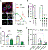
Figure 2.. Inactivation of Inc protein GarD (CTL0390) renders C. trachomatis susceptible to IFNγ-primed host defense.
(A) Immunostaining of endogenous GarD and radial line tracing demonstrates its colocalization with inclusion membrane marker InaC in naïve A549 cells at 20 hpi. Scale bar = 10 μm. (B) IR values reflecting IFNγ-mediated suppression of bacterial growth are shown for garD mutant CTL0390G136E, three independent GII mutants (CTL0390::GII), and their corresponding WT controls. CTL0390::GII#1 was renamed garD::GII and selected for subsequent studies. (C) IFNγ-mediated growth restriction of WT and garD::GII C.t. serovar L2 was measured in HFF-1 and primary cervical epithelial cells using Cellomics high content imaging. (D) Complementation of CTL0390::GII partially restores bacterial growth inside IFNγ-primed A549 cells. Data show the means ± S.D. of three independent experiments. 1-way (B) or 2-way ANOVA (D) followed by Tukey’s multiple comparison test, or an unpaired t-test (C) were performed to assess statistical significance. **= p <0.01, *** = p <0.005, **** = p <0.0001; a.u. = arbitrary units.
Figure 3.
Figure 3.. GarD acts in cis to protect C. trachomatis inclusions from ubiquitylation.
Percentages of inclusions positive for (A) total (FK2) or (B) K48-, K63-, or M-linked (linear) ubiquitin assessed by immunofluorescence in IFNγ-primed (100U/mL) and naïve A549s at 20 hours post infection (hpi). (C) Co-infection of IFNγ-primed (100U/mL) A549s with indicated C.t. strains to distinguish between trans- and cis-acting activity of GarD in blocking inclusion ubiquitylation at 20hpi. Representative confocal images show inclusions within IFNγ-primed cells. Scale bars = 10 μm. Diagram created using BioRender. Data show the means ± S.D. of three independent experiments. 2-way ANOVA followed by Tukey’s multiple comparison test was performed. *= p <0.05, **** = p <0.0001; n.s. = not significant.
Figure 4.
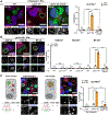
Figure 4.. GarD-deficient inclusions become decorated with ubiquitin adaptor proteins and delivered into LC3+/GABARAPs+ and LAMP1+ compartments.
Immunofluorescence-based quantification of inclusions decorated with (A) ubiquitin-binding proteins OPTN, NDP52, or TAX1BP1, (B) ubiquitin-like LC3 and GABARAP family proteins, and (C) lysosomal LAMP1 in naïve and IFNγ-primed (100U/mL) A549 cells at 20 hours post infection (hpi). Representative confocal images show inclusions in IFNγ-primed cells. Scale bars = 5 μm. Data show the means ± S.D. of at least three independent experiments. 2-way ANOVA followed by Tukey’s multiple comparison test was performed. *= p <0.05, **** = p <0.0001; n.s. = not significant.
Figure 5.
Figure 5.. Human genetic diversity identifies RNF213 as candidate anti-Chlamydia E3-ubiquitin ligase
(A) Stratified QQ plots examining SNPs associated with median C. trachomatis serovar L2 GFP fluorescence at 27 hpi. Empirical P-values were calculated from family-based association analysis using QFAM-parents in PLINK. (B) Regional Manhattan plot around rs12051852 demonstrates that SNPs associated with C. trachomatis burden overlap only the RNF213 gene. SNPs are plotted by position on chromosome 17 and -log(p-value) and color-coded by r2 value to rs12051852 from 1000 Genomes European data. (C) Median C. trachomatis GFP fluorescence in infected cells at 27 hpi plotted by rs12051852 genotype. Each dot represents a single lymphoblastoid cell line (LCL), averaged between three independent experiments. Line marks the median and box indicates the first and third quartiles.
Figure 6.
Figure 6.. GarD protects inclusions against attack by the anti-Chlamydia host ubiquitin E3 ligase RNF213.
Immunostaining demonstrates co-localization of RNF213 with ubiquitin on garD::GII inclusions in (A) A549, (B) HFF-1 and primary cervical epithelial cells at 20 hours post infection (hpi). In (A), radial line tracing of fluorescent intensity is shown for RNF213 and ubiquitin. (C-E) Co-localization with ubiquitin in IFNγ-primed and naïve A549 cells with the adaptor proteins (C) OPTN, (D) NDP52, and (E) TAX1BP1. (F) RNF213 localization to inclusions formed during co-infection of IFNγ-primed and naive A549 cells with garD::GII and non-fusogenic incA or fusogenic WT at 20hpi. (G) IR for WT and garD::GII C.t. in WT and three independent CRISPR-generated RNF213 KO pools at 24 hpi. Representative confocal images show inclusions within IFNγ-primed cells. Scale bars = 10 μm. All data depict the mean ± S.D. from at least three independent experiments. 2-way ANOVA followed by Tukey’s multiple comparison test was used to determine significance. *= p <0.05, **= p <0.01, **** = p <0.0001; n.s. = not significant. (A-E) Statistical comparisons shown for “Both” groups (orange bars).

References

    1. Agaisse H, and Derre I (2013). A C. trachomatis cloning vector and the generation of C. trachomatis strains expressing fluorescent proteins under the control of a C. trachomatis promoter. PLoS One 8, e57090. 10.1371/journal.pone.0057090. - DOI - PMC - PubMed
    1. Ahel J, Lehner A, Vogel A, Schleiffer A, Meinhart A, Haselbach D, and Clausen T (2020). Moyamoya disease factor RNF213 is a giant E3 ligase with a dynein-like core and a distinct ubiquitin-transfer mechanism. Elife 9. 10.7554/eLife.56185. - DOI - PMC - PubMed
    1. Aiyar A, Quayle AJ, Buckner LR, Sherchand SP, Chang TL, Zea AH, Martin DH, and Belland RJ (2014). Influence of the tryptophan-indole-IFNgamma axis on human genital Chlamydia trachomatis infection: role of vaginal co-infections. Front Cell Infect Microbiol 4, 72. 10.3389/fcimb.2014.00072. - DOI - PMC - PubMed
    1. Anand I, Choi W, and Isberg RR (2020). The vacuole guard hypothesis: how intravacuolar pathogens fight to maintain the integrity of their beloved home. Curr Opin Microbiol 54, 51–58. 10.1016/j.mib.2020.01.008. - DOI - PMC - PubMed
    1. Andersen SE, Bulman LM, Steiert B, Faris R, and Weber MM (2021). Got mutants? How advances in chlamydial genetics have furthered the study of effector proteins. Pathog Dis 79. 10.1093/femspd/ftaa078. - DOI - PMC - PubMed