. 2022 Jul:2022:3081-3084.
doi: 10.1109/EMBC48229.2022.9871332.

Demonstration of an Optimized Large-scale Optogenetic Cortical Interface for Non-human Primates

Demonstration of an Optimized Large-scale Optogenetic Cortical Interface for Non-human Primates

Devon J Griggs et al. Annu Int Conf IEEE Eng Med Biol Soc. 2022 Jul.

Abstract

Optogenetics is a powerful neuroscientific tool which allows neurons to be modulated by optical stimulation. Despite widespread optogenetic experimentation in small animal models, optogenetics in non-human primates (NHPs) remains a niche field, particularly at the large scales necessary for multi-regional neural research. We previously published a large-scale, chronic optogenetic cortical interface for NHPs which was successful but came with a number of limitations. In this work, we present an optimized interface which improves upon the stability and scale of our previous interface while using more easily replicable methods to increase our system's availability to the scientific community. Specifically, we (1) demonstrate the long-term (~3 months) optical access to the brain achievable using a commercially-available transparent artificial dura with embedded electrodes, (2) showcase large-scale optogenetic expression achievable with simplified (magnetic resonance-free) surgical techniques, and (3) effectively modulated the expressing areas at large scales (~1 cm2) by light emitting diode (LED) arrays assembled in-house.


Figures

Figure 1:
Figure 1:
Cortical interface. (a) Recreation of NHP skull from MRI data with added craniotomy. (b) Skull-fitting flange of chamber. (c) Chamber fitted on a 3D printed recreation of NHP skull. (d) Stack-up for electrophysiology recording. (e) Stack-up for LED stimulation.
Figure 2:
Figure 2:
LED array. Circuit diagram for LED array and driving electronics (left). Each row of LEDs has its own copy of the driver circuit shown, all sharing a common power supply and occupying the same PCB (schematic, top right; image, bottom right).
Figure 3:
Figure 3:
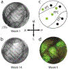
Optical access and optogenetic expression. Image of MMAD on the cortex one week (a) and 14 weeks (b) after implantation and infusion of optogenetic viral vector. (c) Map of optogenetic viral vector injection locations. Green circles indicate successful injections and black circles indicate unsuccessful injections. (d) Epifluorescence imaging of optogenetic expression five weeks after implantation and infusion.
Figure 4:
Figure 4:
Neurophysiology. (a) Time-averaged wideband signals from laser light illumination (30 pulses, 900 ms per pulse at 110 mA, 27 mW optical power) in vivo. The stimulated channel is indicated with an arrow. Stimulation times are shown in red. (b) Time-averaged wideband signals from 30 pulses of the full LED array illumination (30 pulses, 900 ms per pulse at 700 mV, ~2.4 mW/mm2 optical power) in vivo. (c) Averaged power spectral density (PSD) before (top) and during (bottom) LED array stimulation in vivo. (d) Ratio of the PSD (during stim / before stim) for in vivo (top) and saline data (bottom). Dashed line indicates no change. Data above the dashed line indicates excitation, and below the line indicates inhibition. Shaded error bars are 95% confidence interval of median.

References

    1. Yizhar O, Fenno LE, Davidson TJ, Mogri M, and Deisseroth K, “Optogenetics in neural systems,” Neuron, vol. 71, no. 1, pp. 9–34, 2011, doi: 10.1016/j.neuron.2011.06.004. - DOI - PubMed
    1. El-Shamayleh Y and Horwitz GD, “Primate optogenetics: Progress and prognosis,” Proc. Natl. Acad. Sci. U. S. A, vol. 116, no. 52, pp. 26195–26203, 2019, doi: 10.1073/pnas.1902284116. - DOI - PMC - PubMed
    1. Galvan A et al., “Nonhuman primate optogenetics: Recent advances and future directions,” J. Neurosci, vol. 37, no. 45, pp. 10894–10903, 2017, doi: 10.1523/JNEUROSCI.1839-17.2017. - DOI - PMC - PubMed
    1. Galvan A, Caiola MJ, and Albaugh DL, “Advances in optogenetic and chemogenetic methods to study brain circuits in non-human primates,” J Neural Transm, vol. 125, no. 3, pp. 547–563, 2018, doi: 10.1007/s00702-017-1697-8. - DOI - PMC - PubMed
    1. Tremblay S et al., “An Open Resource for Non-human Primate Optogenetics,” Neuron, pp. 1–16, 2020, doi: 10.1016/j.neuron.2020.09.027. - DOI - PMC - PubMed

Publication types

LinkOut - more resources