

Tumour necrosis factor blockade after asphyxia in foetal sheep ameliorates cystic white matter injury
- PMID: 36087304
- PMCID: PMC10115165
- DOI: 10.1093/brain/awac331
Tumour necrosis factor blockade after asphyxia in foetal sheep ameliorates cystic white matter injury
Abstract
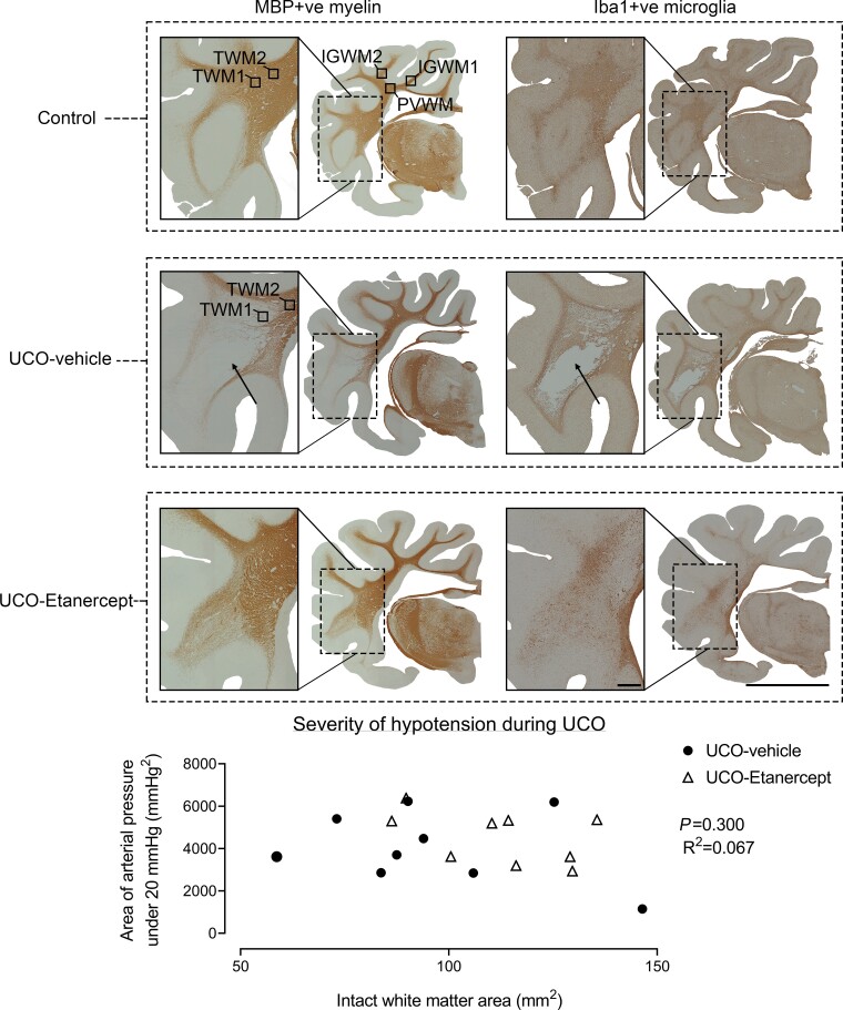
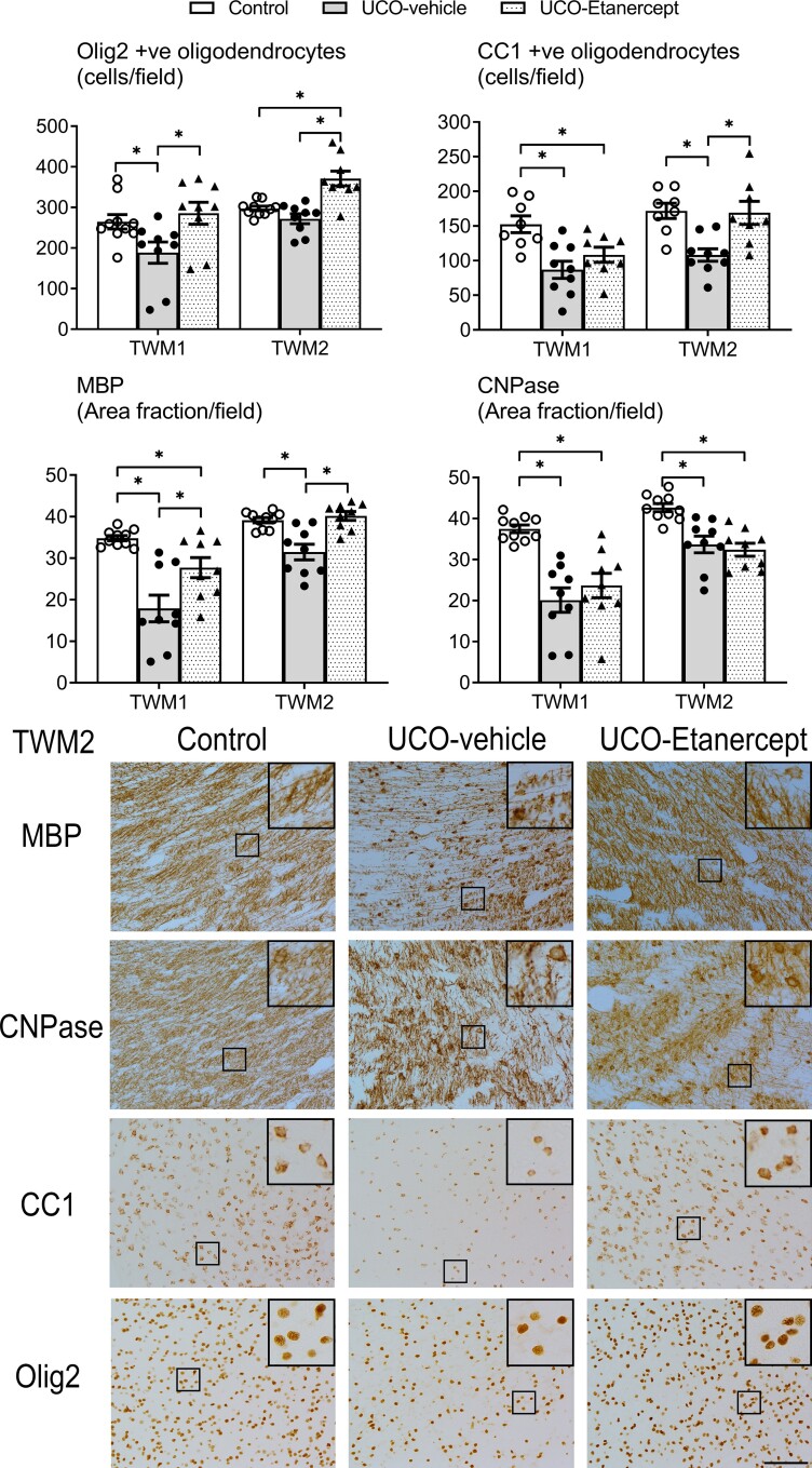
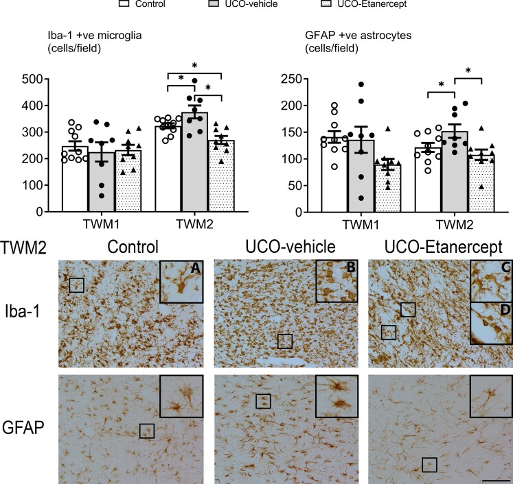
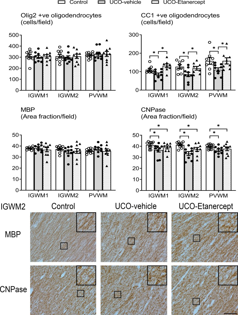
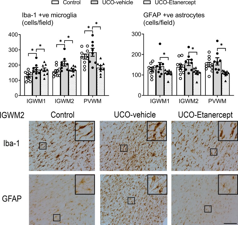
Cystic white matter injury is highly associated with severe neurodevelopmental disability and cerebral palsy in preterm infants, yet its pathogenesis remains poorly understood and there is no established treatment. In the present study, we tested the hypothesis that slowly evolving cystic white matter injury after hypoxia-ischaemia is mediated by programmed necrosis initiated by tumour necrosis factor. Tumour necrosis factor blockade was begun 3 days after hypoxia-ischaemia to target the tertiary phase of injury, when most secondary cell death is thought to be complete. Chronically instrumented preterm foetal sheep (0.7 gestation) received 25 min of hypoxia-ischaemia induced by complete umbilical cord occlusion or sham-umbilical cord occlusion (controls, n = 10), followed by intracerebroventricular infusion of the soluble TNF inhibitor, Etanercept, at 3, 8 and 13 days after umbilical cord occlusion (n = 9) or vehicle (n = 9). Foetal brains were processed for histology at 21 days after umbilical cord occlusion. Umbilical cord occlusion with vehicle was associated with a spectrum of macroscopic white matter degeneration, including white matter atrophy, ventriculomegaly and overt temporal lobe cystic white matter injury. Oligodendrocyte maturational arrest and impaired labelling of myelin proteins, characteristic of diffuse white matter injury, was observed in the parietal lobe and surrounding the cystic lesions in the temporal lobe. Etanercept markedly attenuated cystic white matter injury on the side of the intracerebroventricular infusion, with partial contralateral protection. Further, Etanercept improved oligodendrocyte maturation and labelling of myelin proteins in the temporal and parietal lobes. The present study shows that cystic white matter injury reflects late-onset tertiary cell death mediated by delayed neuroinflammation through the tumour necrosis factor pathway. Delayed tumour necrosis factor blockade markedly attenuated cystic white matter injury and restored oligodendrocyte maturation and deficits in myelin protein expression. These data suggest that delayed tumour necrosis factor blockade may represent a viable therapeutic strategy to reduce the risk of cystic and diffuse white matter injury and potentially cerebral palsy after preterm birth, with a surprisingly wide therapeutic window.
Keywords: Etanercept; hypoxia-ischaemia; periventricular leukomalacia; preterm; white matter injury.
© The Author(s) 2022. Published by Oxford University Press on behalf of the Guarantors of Brain.
Conflict of interest statement
The authors report no competing interests.
Figures
Similar articles
-
Tertiary cystic white matter injury as a potential phenomenon after hypoxia-ischaemia in preterm f sheep.Brain Commun. 2021 Mar 9;3(2):fcab024. doi: 10.1093/braincomms/fcab024. eCollection 2021. Brain Commun. 2021. PMID: 33937767 Free PMC article.
-
White matter protection with insulin-like growth factor-1 after hypoxia-ischaemia in preterm foetal sheep.Brain Commun. 2024 Oct 24;6(6):fcae373. doi: 10.1093/braincomms/fcae373. eCollection 2024. Brain Commun. 2024. PMID: 39507274 Free PMC article.
-
Magnesium sulphate reduces tertiary gliosis but does not improve EEG recovery or white or grey matter cell survival after asphyxia in preterm fetal sheep.J Physiol. 2023 May;601(10):1999-2016. doi: 10.1113/JP284381. Epub 2023 Apr 13. J Physiol. 2023. PMID: 36999348 Free PMC article.
-
Is Late Prevention of Cerebral Palsy in Extremely Preterm Infants Plausible?Dev Neurosci. 2022;44(4-5):177-185. doi: 10.1159/000521618. Epub 2021 Dec 22. Dev Neurosci. 2022. PMID: 34937030 Review.
-
Role of instrumented fetal sheep preparations in defining the pathogenesis of human periventricular white-matter injury.J Child Neurol. 2006 Jul;21(7):582-9. doi: 10.1177/08830738060210070101. J Child Neurol. 2006. PMID: 16970848 Review.
Cited by
-
Dysmaturation of sleep state and electroencephalographic activity after hypoxia-ischaemia in preterm fetal sheep.J Cereb Blood Flow Metab. 2024 Aug;44(8):1376-1392. doi: 10.1177/0271678X241236014. Epub 2024 Feb 28. J Cereb Blood Flow Metab. 2024. PMID: 38415649 Free PMC article.
-
A systematic review of immune-based interventions for perinatal neuroprotection: closing the gap between animal studies and human trials.J Neuroinflammation. 2023 Oct 20;20(1):241. doi: 10.1186/s12974-023-02911-w. J Neuroinflammation. 2023. PMID: 37864272 Free PMC article.
-
Systemic Inflammatory Response Syndrome, Thromboinflammation, and Septic Shock in Fetuses and Neonates.Int J Mol Sci. 2025 Apr 1;26(7):3259. doi: 10.3390/ijms26073259. Int J Mol Sci. 2025. PMID: 40244141 Free PMC article. Review.
-
Circadian patterns of heart rate variability in fetal sheep after hypoxia-ischaemia: A biomarker of evolving brain injury.J Physiol. 2024 Dec;602(23):6553-6569. doi: 10.1113/JP284560. Epub 2023 Jul 11. J Physiol. 2024. PMID: 37432936 Free PMC article.
-
Oligodendrocyte Progenitor Cell Transplantation Reduces White Matter Injury in a Fetal Goat Model.CNS Neurosci Ther. 2024 Dec;30(12):e70178. doi: 10.1111/cns.70178. CNS Neurosci Ther. 2024. PMID: 39690788 Free PMC article.
References
-
- Galea C, McIntyre S, Smithers-Sheedy H, et al. . Cerebral palsy trends in Australia (1995–2009): A population-based observational study. Dev Med Child Neurol. 2019;61:186–193. - PubMed
-
- Ghotra S, Vincer M, Allen VM, Khan N. A population-based study of cystic white matter injury on ultrasound in very preterm infants born over two decades in Nova Scotia, Canada. J Perinatol. 2019;39:269–277. - PubMed
-
- Banker BQ, Larroche JC. Periventricular leukomalacia of infancy. A form of neonatal anoxic encephalopathy. Arch Neurol. 1962;7:386–410. - PubMed
-
- Hamrick SE, Miller SP, Leonard C, et al. . Trends in severe brain injury and neurodevelopmental outcome in premature newborn infants: The role of cystic periventricular leukomalacia. J Pediatr. 2004;145:593–599. - PubMed
