

Breast dosimetry in alternative X-ray-based imaging modalities used in current clinical practices
- PMID: 36087425
- PMCID: PMC9851082
- DOI: 10.1016/j.ejrad.2022.110509
Breast dosimetry in alternative X-ray-based imaging modalities used in current clinical practices
Abstract
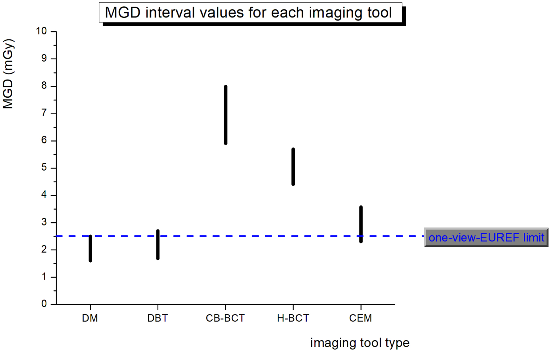
In X-ray breast imaging, Digital Mammography (DM) and Digital Breast Tomosynthesis (DBT), are the standard and largely used techniques, both for diagnostic and screening purposes. Other techniques, such as dedicated Breast Computed Tomography (BCT) and Contrast Enhanced Mammography (CEM) have been developed as an alternative or a complementary technique to the established ones. The performance of these imaging techniques is being continuously assessed to improve the image quality and to reduce the radiation dose. These imaging modalities are predominantly used in the diagnostic setting to resolve incomplete or indeterminate findings detected with conventional screening examinations and could potentially be used either as an adjunct or as a primary screening tool in select populations, such as for women with dense breasts. The aim of this review is to describe the radiation dosimetry for these imaging techniques, and to compare the mean glandular dose with standard breast imaging modalities, such as DM and DBT.
Keywords: Breast Computed Tomography; Breast dosimetry; Contrast Enhanced Mammography; Mean Glandular Dose.
Copyright © 2022 Elsevier B.V. All rights reserved.
Conflict of interest statement
Declaration of Competing Interest The authors declare that they have no known competing financial interests or personal relationships that could have appeared to influence the work reported in this paper.
Figures


References
-
- Osteras BH, Martinsen ACT, Gullien R and Skaane P, “Digital Mammography versus Breast Tomosynthesis: Impact of Breast Density on Diagnostic Performance in Population-based Screening,” Radiology, pp. 60–68, 2019. - PubMed
-
- Skaane P, Bandos AI, Niklason LT, Sebuødegård S, Østerås BH, Gullien R, Gur D and Hofvind S, “Digital Mammography versus Digital Mammography Plus Tomosynthesis in Breast Cancer Screening: The Oslo Tomosynthesis Screening Trial,” Radiology, vol. 291, no. 1, pp. 23–30, Apr 2019. DOI: 10.1148/radiol.2019182394. - DOI - PubMed
Publication types
MeSH terms
Grants and funding
LinkOut - more resources
Full Text Sources
Medical
