

Recommendations on compiling test datasets for evaluating artificial intelligence solutions in pathology
- PMID: 36088478
- PMCID: PMC9708586
- DOI: 10.1038/s41379-022-01147-y
Recommendations on compiling test datasets for evaluating artificial intelligence solutions in pathology
Erratum in
-
Publisher Correction to: Recommendations on compiling test datasets for evaluating artificial intelligence solutions in pathology.Mod Pathol. 2022 Dec;35(12):2034. doi: 10.1038/s41379-022-01163-y. Mod Pathol. 2022. PMID: 36151301 Free PMC article. No abstract available.
Abstract
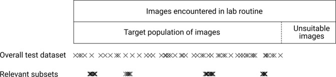
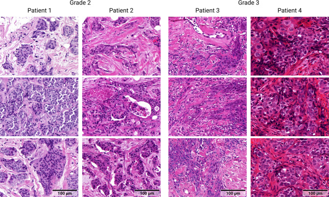
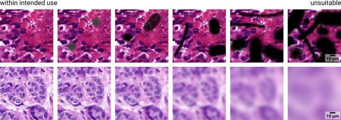
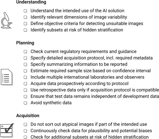
Artificial intelligence (AI) solutions that automatically extract information from digital histology images have shown great promise for improving pathological diagnosis. Prior to routine use, it is important to evaluate their predictive performance and obtain regulatory approval. This assessment requires appropriate test datasets. However, compiling such datasets is challenging and specific recommendations are missing. A committee of various stakeholders, including commercial AI developers, pathologists, and researchers, discussed key aspects and conducted extensive literature reviews on test datasets in pathology. Here, we summarize the results and derive general recommendations on compiling test datasets. We address several questions: Which and how many images are needed? How to deal with low-prevalence subsets? How can potential bias be detected? How should datasets be reported? What are the regulatory requirements in different countries? The recommendations are intended to help AI developers demonstrate the utility of their products and to help pathologists and regulatory agencies verify reported performance measures. Further research is needed to formulate criteria for sufficiently representative test datasets so that AI solutions can operate with less user intervention and better support diagnostic workflows in the future.
© 2022. The Author(s).
Conflict of interest statement
F.Z. is a shareholder of asgen GmbH. P.S. is a member of the supervisory board of asgen GmbH. All other authors declare that they have no conflict of interest.
Figures
References
-
- Moxley-Wyles B, Colling R, Verrill C. Artificial intelligence in pathology: An overview. Diagn Histopathol 26, 513–520 (2020).
Publication types
MeSH terms
LinkOut - more resources
Full Text Sources
