Review
. 2022 Oct:141:104855.
doi: 10.1016/j.neubiorev.2022.104855. Epub 2022 Sep 8.

Neuronal circuitry for recognition memory of object and place in rodent models

Affiliations
Review

Neuronal circuitry for recognition memory of object and place in rodent models

Owen Y Chao et al. Neurosci Biobehav Rev. 2022 Oct.

Abstract

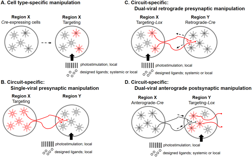
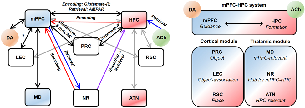
Rats and mice are used for studying neuronal circuits underlying recognition memory due to their ability to spontaneously remember the occurrence of an object, its place and an association of the object and place in a particular environment. A joint employment of lesions, pharmacological interventions, optogenetics and chemogenetics is constantly expanding our knowledge of the neural basis for recognition memory of object, place, and their association. In this review, we summarize current studies on recognition memory in rodents with a focus on the novel object preference, novel location preference and object-in-place paradigms. The evidence suggests that the medial prefrontal cortex- and hippocampus-connected circuits contribute to recognition memory for object and place. Under certain conditions, the striatum, medial septum, amygdala, locus coeruleus and cerebellum are also involved. We propose that the neuronal circuitry for recognition memory of object and place is hierarchically connected and constructed by different cortical (perirhinal, entorhinal and retrosplenial cortices), thalamic (nucleus reuniens, mediodorsal and anterior thalamic nuclei) and primeval (hypothalamus and interpeduncular nucleus) modules interacting with the medial prefrontal cortex and hippocampus.

Keywords: Cell type specificity; Entorhinal cortex; Hippocampus; Medial prefrontal cortex; Object recognition; Spatial memory; Thalamus.


Conflict of interest statement

Declaration of Competing Interest The authors declare that they have no known competing financial interests or personal relationships that could have appeared to influence the work reported in this paper.

Figures

Figure 1.
Figure 1.
Schematic diagrams of spontaneous object exploration paradigms of the novel object preference (NOP), novel location preference (NLP) and object-in-place preference (OiP). Circled objects are explored more than non-circled objects by rodents in the testing trial.
Figure 2.
Figure 2.
Strategies for viral vector-based genetic manipulations of neuronal activities. (A) Neurons expressing Cre (solid-gray dots) are targeted by a viral vector carrying DIO (red). Via the Cre-Lox recombination (as an example), this design enables bidirectional manipulation of a specific neuronal population in a brain region with optogenetic or chemogenetic approaches. (B) Neurons in source region X are targeted by a viral vector. Their axonal terminals in downstream region Y can be manipulated with optogenetic or chemogenetic stimulations. (C) A dual-viral retrograde targeting method to manipulate presynaptic neurons. A retrograde virus carrying Cre is injected into region Y, while another viral vector carrying DIO is injected into upstream region X. Via the Cre-Lox recombination, only neurons, which project to the Cre-injected region, are selectively manipulated. (D) A dual-viral anterograde targeting method to manipulate postsynaptic neurons. An anterograde virus carrying Cre is injected into region X, while another viral vector carrying DIO is injected into downstream region Y. Via the Cre-Lox recombination, only neurons, which receive projections from the Cre-injected region, are selectively manipulated. Neuronal manipulations can be achieved by photostimulation (optogenetics) or designed ligands (chemogenetics).
Figure 3.
Figure 3.
Common methods for studying brain function are classified by their specificity (X-axis) and temporal (Y-axis) profiles. In terms of specificity, viral-based genetic methods can target a specific cell type or circuit (blue fonts), while other methods are limited (neurotoxins can target a certain type of neurons but are difficult to apply for specific circuits). In contrast to chronic manipulations, transient manipulations offer high temporal precision and are reversible. They may, however, incur “off-target” effects (green background). DREADDs: designer receptors exclusively activated by designer drugs; DTA: diphtheria toxin subunit A; tDCS: transcranial direct current stimulation; TeLC: tetanus toxin light chain; TMS: transcranial magnetic stimulation.
Figure 4.
Figure 4.
Neuroanatomy of recognition memory based on spontaneous object exploration tests in rodents. Blue and red lines show the mPFC- and HPC-connected circuits, respectively. Black lines show the projections sent from the LC and cerebellum to the dorsal HPC and thalamus, respectively. Note that other regions (IC, striatum, MS, hypothalamus, amygdala and IPN) are highly connected with the mentioned structures and contribute to recognition memory under certain conditions. The right panel shows a hypothetical model of recognition memory based on hierarchically organized, functionally distinct, yet complementary anatomical connectivity: Hypothalamus and IPN send information of familiarity and novelty (F/N signals) to the thalamic nuclei. The thalamic nuclei, as integrators, organize the incoming F/N signals and actively interact with the mPFC-HPC system and cortices. The HPC generates essential mnemonic information featured with spatial and contextual information, while the mPFC selects the “correct” memory dependent on environmental requirements; both interact with the reciprocally connected cortices that support the mPFC-HPC system. The mPFC-MD (blue) and HPC-ATN (red) circuits preferentially modulate information for memory guidance and formation, respectively, gated by the NR. Dashed lines represent possible connectivity. * The pathway from the mPFC to the HPC is debated (see Andrianova et al., 2022). mPFC: medial prefrontal cortex; HPC: hippocampus; RSC: retrosplenial cortex; PRC: perirhinal cortex; EC: entorhinal cortex (the lateral EC mutually connects with the mPFC and HPC); IC: insular cortex; MS: medial septum; IPN: interpeduncular nucleus; LC: locus coeruleus; MD: mediodorsal thalamus; NR: nucleus reuniens; ATN: anterior thalamic nucleus.
Figure 5.
Figure 5.
A hypothetical circuit of the associative memory for object-place. Arrow lines indicate that the interaction between the linked regions is essential for object-place association memory. The connections are color-coded for either encoding (red), retrieval (blue) or encoding plus retrieval (purple). Gray lines indicate functional interactions between the hypothetically linked regions, which, so far, have not been tested. * The interaction between the mPFC and LEC has not been directly examined in the object-in-place memory test; however, this pathway is likely to contribute to the association of object and place. Note that the NR functional circuits are mainly based on a preprint paper (Barker et al., 2021). The right panel depicts the mPFC-HPC system responsible for memory guidance and formation, the cortical module accounting for object (PRC), object-association (LEC) and place (RSC) information, and the thalamic module processing the mPFC-relevant (MD), HPC-relevant (ATN) and mPFC-HPC messages (NR), which can be modulated by DA and ACh (presumably via the medial septum connections). mPFC: medial prefrontal cortex; HPC: hippocampus; LEC: lateral entorhinal cortex; PRC: perirhinal cortex; RSC: retrosplenial cortex; MD: mediodorsal thalamus; NR: nucleus reuniens; ATN: anterior thalamus; DA: dopamine; ACh: acetylcholine; glutamate-R: glutamate receptor; mAChR: muscarinic acetylcholine receptor; AMPAR: AMPA receptor.

References

    1. Aarse J, Herlitze S, Manahan-Vaughan D (2016) The requirement of BDNF for hippocampal synaptic plasticity is experience-dependent. Hippocampus 26:739–751. - PMC - PubMed
    1. Abe H, Ishida Y, Nonaka H, Iwasaki T (2009) Functional difference between rat perirhinal cortex and hippocampus in object and place discrimination tasks. Behav Brain Res 197:388–397. - PubMed
    1. Adamantidis A, de Lecea L (2009) A role for melanin-concentrating hormone in learning and memory. Peptides 30:2066–2070. - PMC - PubMed
    1. Aggleton JP (1985) One-trial object recognition by rats. Q J Exp Psychol-B 37:279–294.
    1. Aggleton JP (2010) Understanding retrosplenial amnesia: insights from animal studies. Neuropsychologia 48:2328–2338. - PubMed

Publication types