

Neuronal cell death mechanisms in Alzheimer's disease: An insight
- PMID: 36090249
- PMCID: PMC9454331
- DOI: 10.3389/fnmol.2022.937133
Neuronal cell death mechanisms in Alzheimer's disease: An insight
Abstract
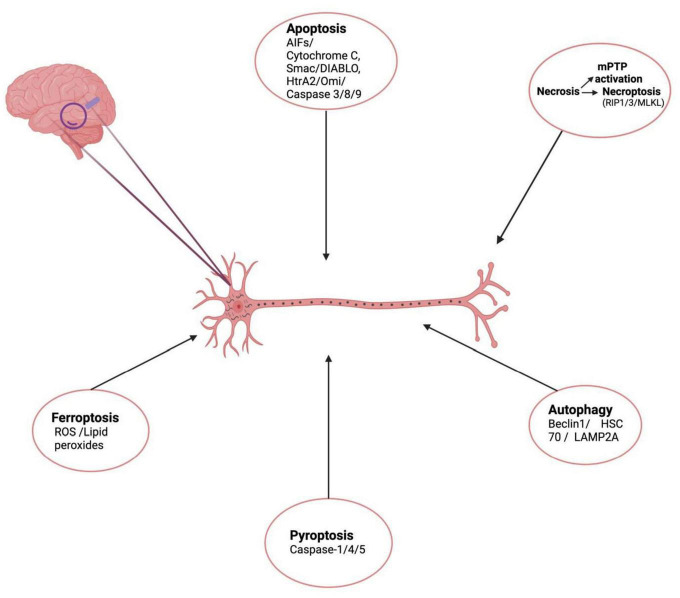
Regulated cell death (RCD) is an ordered and tightly orchestrated set of changes/signaling events in both gene expression and protein activity and is responsible for normal development as well as maintenance of tissue homeostasis. Aberrant activation of this pathway results in cell death by various mechanisms including apoptosis, necroptosis, pyroptosis, ferroptosis, and autophagy-dependent cell death. Such pathological changes in neurons alone or in combination have been observed in the pathogenesis of various neurodegenerative diseases including Alzheimer's disease (AD). Pathological hallmarks of AD focus primarily on the accumulation of two main protein markers: amyloid β peptides and abnormally phosphorylated tau proteins. These protein aggregates result in the formation of A-β plaques and neuro-fibrillary tangles (NFTs) and induce neuroinflammation and neurodegeneration over years to decades leading to a multitude of cognitive and behavioral deficits. Autopsy findings of AD reveal massive neuronal death manifested in the form of cortical volume shrinkage, reduction in sizes of gyri to up to 50% and an increase in the sizes of sulci. Multiple forms of cell death have been recorded in neurons from different studies conducted so far. However, understanding the mechanism/s of neuronal cell death in AD patients remains a mystery as the trigger that results in aberrant activation of RCD is unknown and because of the limited availability of dying neurons. This review attempts to elucidate the process of Regulated cell death, how it gets unregulated in response to different intra and extracellular stressors, various forms of unregulated cell death, their interplay and their role in pathogenesis of Alzheimer's Disease in both human and experimental models of AD. Further we plan to explore the correlation of both amyloid-beta and Tau with neuronal loss as seen in AD.
Keywords: Alzheimer’s disease; apoptosis; autophagy; ferroptosis; necroptosis.
Copyright © 2022 Goel, Chakrabarti, Goel, Bhutani, Chopra and Bali.
Conflict of interest statement
The authors declare that the research was conducted in the absence of any commercial or financial relationships that could be construed as a potential conflict of interest.
Figures
References
-
- Albrecht S., Bogdanovic N., Ghetti B., Winblad B., LeBlanc A. C. (2009). Caspase-6 activation in familial Alzheimer disease brains carrying amyloid precursor protein or presenilin I or presenilin II mutations. J. Neuropathol. Exp. Neurol. 68 1282–1293. 10.1097/NEN.0b013e3181c1da10 - DOI - PMC - PubMed
Publication types
LinkOut - more resources
Full Text Sources
Other Literature Sources