Different Features of 18F-FAPI, 18F-FDG PET/CT and MRI in the Evaluation of Extrahepatic Metastases and Local Recurrent Hepatocellular Carcinoma (HCC): A Case Report and Review of the Literature
- PMID: 36090470
- PMCID: PMC9462837
- DOI: 10.2147/CMAR.S374916
Different Features of 18F-FAPI, 18F-FDG PET/CT and MRI in the Evaluation of Extrahepatic Metastases and Local Recurrent Hepatocellular Carcinoma (HCC): A Case Report and Review of the Literature
Abstract
Background: Recurrence and metastasis are important causes of postoperative death in most HCC patients. Conventional imaging modalities such as 18F-FDG PET/CT and enhanced MRI are still unsatisfactory in evaluating these patients in the clinical setting. PET/CT imaging with a radiolabeled fibroblast activation protein inhibitor (FAPI) has emerged as a new imaging technique for the diagnosis and radiotherapy of malignant tumors. While many studies have focused on the diagnostic accuracy of intrahepatic primary HCC, the evaluation of recurrent and metastatic HCC remains only poorly investigated.
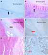
Case presentation: A 71-year-old man with a five-year history of HCC after radical resection underwent 18F-FDG PET/CT due to further surgery for tumor recurrence, which revealed two iso-metabolic lesions in the right peritoneum and a hypo-metabolic lesion in the right liver. 18F-FAPI PET/CT was performed to further complement 18F-FDG PET/CT in the detection of these suspected metastatic lesions. Importantly, multiple diffuse intense radioactivity was shown in the hepatic capsule, suggesting metastatic lesions, but a wedge-shaped elevated 18F-FAPI uptake disorder around the FDG-unavid necrotic lesion after radiofrequency ablation (RFA) demonstrated benign stromal fibrosis.
Conclusion: This case suggested that 18F-FAPI may have an advantage over 18F-FDG in detecting peritoneal metastasis even in tiny or early hepatic capsules of HCC, but its false positives due to postoperative stromal fibrosis should be noted. Wedge- or strip-shaped FAPI-avid lesions with sharp edges may be post-treatment stromal fibrosis.
Keywords: 18F-FAPI; 18F-FDG; PET/CT; false-positive; hepatocellular carcinoma; magnetic resonance imaging; stromal fibrosis.
© 2022 Chen et al.
Conflict of interest statement
The authors declare that they have no competing interests.
Figures


Similar articles
-
Comparison of PET imaging of activated fibroblasts and 18F-FDG for diagnosis of primary hepatic tumours: a prospective pilot study.Eur J Nucl Med Mol Imaging. 2021 May;48(5):1593-1603. doi: 10.1007/s00259-020-05070-9. Epub 2020 Oct 24. Eur J Nucl Med Mol Imaging. 2021. PMID: 33097975
-
Imaging fibroblast activation protein in liver cancer: a single-center post hoc retrospective analysis to compare [68Ga]Ga-FAPI-04 PET/CT versus MRI and [18F]-FDG PET/CT.Eur J Nucl Med Mol Imaging. 2021 May;48(5):1604-1617. doi: 10.1007/s00259-020-05095-0. Epub 2020 Nov 11. Eur J Nucl Med Mol Imaging. 2021. PMID: 33179149
-
[18F]FAPI PET/CT in the evaluation of focal liver lesions with [18F]FDG non-avidity.Eur J Nucl Med Mol Imaging. 2023 Feb;50(3):937-950. doi: 10.1007/s00259-022-06022-1. Epub 2022 Nov 8. Eur J Nucl Med Mol Imaging. 2023. PMID: 36346437
-
Comparison of 68Ga-FAPI and 18F-FDG PET/CT for the diagnosis of primary and metastatic lesions in abdominal and pelvic malignancies: A systematic review and meta-analysis.Front Oncol. 2023 Feb 17;13:1093861. doi: 10.3389/fonc.2023.1093861. eCollection 2023. Front Oncol. 2023. PMID: 36874127 Free PMC article.
-
FAPI PET/CT in the Diagnosis of Abdominal and Pelvic Tumors.Front Oncol. 2022 Jan 4;11:797960. doi: 10.3389/fonc.2021.797960. eCollection 2021. Front Oncol. 2022. PMID: 35059319 Free PMC article. Review.
Cited by
-
Diagnostic Performances of PET/CT Using Fibroblast Activation Protein Inhibitors in Patients with Primary and Metastatic Liver Tumors: A Comprehensive Literature Review.Int J Mol Sci. 2024 Jun 29;25(13):7197. doi: 10.3390/ijms25137197. Int J Mol Sci. 2024. PMID: 39000301 Free PMC article. Review.
-
Assessment of viable tumours by [68Ga]Ga-FAPI-04 PET/CT after local regional treatment in patients with hepatocellular carcinoma.Eur J Nucl Med Mol Imaging. 2025 May;52(6):2132-2144. doi: 10.1007/s00259-024-07062-5. Epub 2025 Jan 20. Eur J Nucl Med Mol Imaging. 2025. PMID: 39831967
-
18F-5-fluoro-aminosuberic acid PET/CT imaging of oxidative-stress features during the formation of DEN-induced rat hepatocellular carcinoma.Theranostics. 2025 Jan 1;15(1):141-154. doi: 10.7150/thno.100467. eCollection 2025. Theranostics. 2025. PMID: 39744233 Free PMC article.
References
Publication types
LinkOut - more resources
Full Text Sources

