. 2022 Aug 25:16:953157.
doi: 10.3389/fnbeh.2022.953157. eCollection 2022.

Palmitoylethanolamide attenuates neurodevelopmental delay and early hippocampal damage following perinatal asphyxia in rats

Affiliations

Palmitoylethanolamide attenuates neurodevelopmental delay and early hippocampal damage following perinatal asphyxia in rats

Maria I Herrera et al. Front Behav Neurosci. .

Erratum in

Abstract

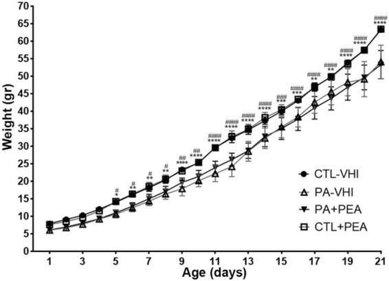
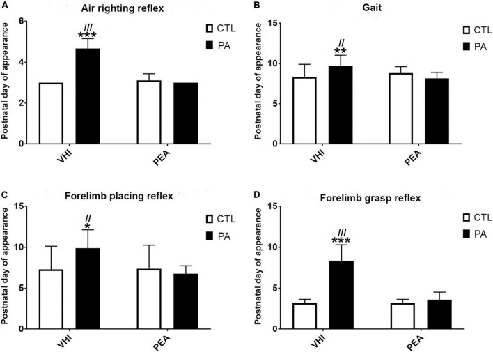
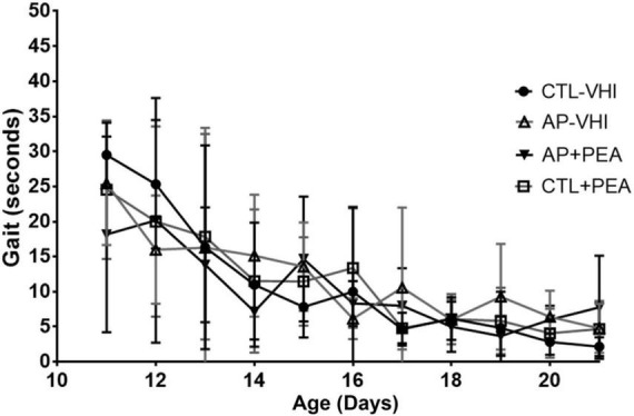
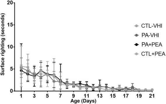
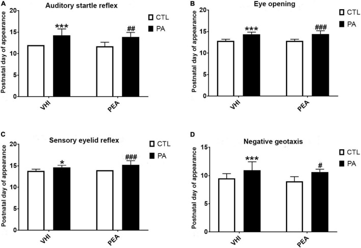
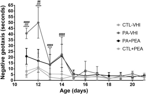
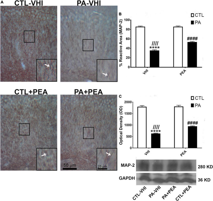
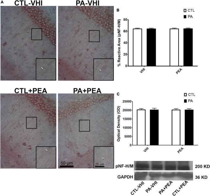
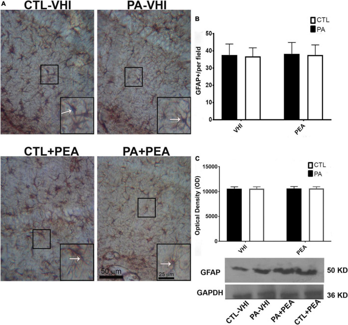
Impaired gas exchange close to labor causes perinatal asphyxia (PA), a neurodevelopmental impairment factor. Palmitoylethanolamide (PEA) proved neuroprotective in experimental brain injury and neurodegeneration models. This study aimed to evaluate PEA effects on the immature-brain, i.e., early neuroprotection by PEA in an experimental PA paradigm. Newborn rats were placed in a 37°C water bath for 19 min to induce PA. PEA 10 mg/kg, s.c., was administered within the first hour of life. Neurobehavioral responses were assessed from postnatal day 1 (P1) to postnatal day 21 (P21), recording the day of appearance of several reflexes and neurological signs. Hippocampal CA1 area ultrastructure was examined using electron microscopy. Microtubule-associated protein 2 (MAP-2), phosphorylated high and medium molecular weight neurofilaments (pNF H/M), and glial fibrillary acidic protein (GFAP) were assessed using immunohistochemistry and Western blot at P21. Over the first 3 weeks of life, PA rats showed late gait, negative geotaxis and eye-opening onset, and delayed appearance of air-righting, auditory startle, sensory eyelid, forelimb placing, and grasp reflexes. On P21, the hippocampal CA1 area showed signs of neuronal degeneration and MAP-2 deficit. PEA treatment reduced PA-induced hippocampal damage and normalized the time of appearance of gait, air-righting, placing, and grasp reflexes. The outcome of this study might prove useful in designing intervention strategies to reduce early neurodevelopmental delay following PA.

Keywords: PEA; hippocampal CA1 area; neurodevelopmental disorder (NDD); neuroprotection; palmitoylethanolamide; perinatal asphyxia; reflexes.


Conflict of interest statement

The authors declare that the research was conducted in the absence of any commercial or financial relationships that could be construed as a potential conflict of interest.

Figures

FIGURE 1
FIGURE 1
Body weight gain. Results are expressed as mean ± SEM. *p < 0.05, PA-VHI vs. CTL-VHI; **p < 0.01, PA-VHI vs. CTL-VHI; ***p < 0.001, PA-VHI vs. CTL-VHI; ****p < 0.0001, PA-VHI vs. CTL-VHI; #p < 0.05, PA + PEA vs. CTL-VHI; ##p < 0.01, PA + PEA vs. CTL-VHI; ###p < 0.001, PA + PEA vs. CTL-VHI; ####p < 0.0001, PA + PEA vs. CTL-VHI. CTL-VHI, control rats treated with vehicle; PA-VHI, rats subjected to PA and treated with vehicle; PA + PEA, rats subjected to PA and treated with PEA; CTL + PEA, control rats treated with PEA. ***p < 0.001 PA-VHI vs. CTL-VHI.
FIGURE 2
FIGURE 2
Neurobehavioral development. (A) Air-righting reflex. (B) Gait. (C) Forelimb placing reflex. (D) Forelimb grasp reflex. Results are expressed as mean ± SEM. *p < 0.05, PA-VHI vs. CTL-VHI; **p < 0.01, PA-VHI vs. CTL-VHI; ***p < 0.001, PA-VHI vs. CTL-VHI; //p < 0.01, PA-VHI vs. PA + PEA; ///p < 0.001, PA-VHI vs. PA + PEA. CTL-VHI, control rats treated with vehicle; PA-VHI, rats subjected to PA and treated with vehicle; PA + PEA, rats subjected to PA and treated with PEA; CTL + PEA, control rats treated with PEA.
FIGURE 3
FIGURE 3
Gait performance. Results are expressed as mean ± SEM. CTL-VHI, control rats treated with vehicle; PA-VHI, rats subjected to PA and treated with vehicle; PA + PEA, rats subjected to PA and treated with PEA; CTL + PEA, control rats treated with PEA.
FIGURE 4
FIGURE 4
Surface righting performance. Results are expressed as mean ± SEM. Statistical analyses were conducted by two-way mixed ANOVA. CTL-VHI, control rats treated with vehicle; PA-VHI, rats subjected to PA and treated with vehicle; PA + PEA, rats subjected to PA and PEA treatment; CTL + PEA, control rats treated with PEA.
FIGURE 5
FIGURE 5
Postnatal day of reflexes appearance and neurological signs. (A) Auditory start reflex. (B) Eye opening. (C) Sensory eyelid reflex. (D) Negative Geotaxis. Bars and error bars represent mean ± SEM. *p < 0.05, PA-VHI vs. CTL-VHI; ***p < 0.001, PA-VHI vs. CTL-VHI; #p < 0.05, PA + PEA vs. CTL-VHI; ##p < 0.01, PA + PEA vs. CTL-VHI; ###p < 0.001, PA + PEA vs. CTL-VHI. CTL-VHI, control rats treated with vehicle; PA-VHI, rats subjected to PA and treated with vehicle; PA + PEA, rats subjected to PA and treated with PEA; CTL + PEA, control rats treated with PEA.
FIGURE 6
FIGURE 6
Negative geotaxis performance. Results are expressed as mean ± SEM. ***p < 0.001, PA-VHI vs. CTL-VHI; ****p < 0.0001, PA-VHI vs. CTL-VHI; ////p < 0.0001, PA-VHI vs. PA + PEA; ##p < 0.01, PA + PEA vs. CTL-VHI; ####p < 0.0001, PA + PEA vs. CTL-VHI. CTL-VHI, control rats treated with vehicle; PA-VHI, rats subjected to PA and treated with vehicle; PA + PEA, rats subjected to PA and treated with PEA; CTL + PEA, control rats treated with PEA.
FIGURE 7
FIGURE 7
Microtubule-associated protein (MAP-2) immunostaining and protein expression in the rat hippocampus on P21. (A) Representative images of the stratum radiatum of CA1 hippocampal area immunostained for MAP-2. Scale bar: 50 μm. Marked rectangular areas in the main-image are shown magnified in the corresponding lower right margin. The white arrow indicates positive immunostaining for MAP-2. Scale bar: 25 μm. (B) Percentage of reactive area of MAP-2 positive dendrites. (C) Optical density of bands showing MAP-2 protein expression in Western blot. Results are shown as mean ± SEM. ****p < 0.0001, PA-VHI vs. CTL-VHI; ////p < 0.0001, PA-VHI vs. PA + PEA; ####p < 0.0001, PA + PEA vs. CTL-VHI. CTL-VHI, control rats treated with vehicle; PA-VHI, rats subjected to PA and treated with vehicle; PA + PEA, rats subjected to PA and treated with PEA; CTL + PEA, control rats treated with PEA.
FIGURE 8
FIGURE 8
Phosphorylated high and medium molecular weight neurofilaments (pNF H/M) immunostaining and protein expression in the rat hippocampus on P21. (A) Representative images of the stratum radiatum of CA1 hippocampal area immunostained for pNF H/M. Scale bar: 50 μm. Marked rectangular areas in the main-image are shown magnified in the corresponding lower right margin. The white arrow indicates positive immunostaining for pNF H/M. Scale bar: 25 μm. (B) Percentage of reactive area of pNF H/M positive axons. (C) Optical density of bands showing pNF H/M protein expression in Western blot. Results are shown as mean ± SEM. PA-VHI, rats subjected to PA and treated with vehicle; PA + PEA, rats subjected to PA and treated with PEA; CTL + PEA, control rats treated with PEA.
FIGURE 9
FIGURE 9
Glial fibrillary acidic protein (GFAP) immunostaining and protein expression in the rat hippocampus on P21. (A) Representative images of the stratum radiatum of CA1 hippocampal area immunostained for GFAP. Scale bar: 50 μm. Marked rectangular areas in the main-image are shown magnified in the corresponding lower right margin. The white arrow indicates positive immunostaining area for GFAP. Scale bar: 25 μm. (B) Percentage of reactive area of GFAP positive astrocytes. (C) Optical density of bands showing GFAP protein expression in Western blot. Results are shown as mean ± SEM. CTL-VHI, control rats treated with vehicle; PA-VHI, rats subjected to PA and treated with vehicle; PA + PEA, rats subjected to PA and treated with PEA; CTL + PEA, control rats treated with PEA.

Similar articles

References

    1. Adcock L. M., Papile L. A. (2008). “Perinatal asphyxia,” in Manual of neonatal care, eds Cloherty J. P., Eichenwald E. C., Stark A. R. (Philadelphia, PA: Lippincott, Williams, and Wilkins; ), 518–528.
    1. Ahmad A., Crupi R., Impellizzeri D., Campolo M., Marino A., Esposito E., et al. (2012). Administration of palmitoylethanolamide (PEA) protects the neurovascular unit and reduces secondary injury after traumatic brain injury in mice. Brain Behav. Immun. 26 1310–1321. 10.1016/j.bbi.2012.07.021 - DOI - PubMed
    1. Antonucci N., Cirillo A., Siniscalco D. (2015). Beneficial Effects of Palmitoylethanolamide on expressive language, cognition, and behaviors in Autism: a report of two cases. Case Rep. Psychiatry 2015:325061. 10.1155/2015/325061 - DOI - PMC - PubMed
    1. Ashworth S. L., Southgate E. L., Sandoval R. M., Meberg P. J., Bamburg J. R., Molitoris B. A. (2003). ADF/cofilin mediates actin cytoskeletal alterations in LLC-PK cells during ATP depletion. Am. J. Physiol. Renal Physiol. 284 F852–F862. 10.1152/ajprenal.00210.2002 - DOI - PubMed
    1. Azzopardi D., Robertson N. J., Bainbridge A., Cady E., Charles-Edwards G., Deierl A., et al. (2016). Moderate hypothermia within 6 h of birth plus inhaled xenon versus moderate hypothermia alone after birth asphyxia (TOBY-Xe): a proof-of-concept, open-label, randomised controlled trial. Lancet Neurol. 15 145–153. 10.1016/S1474-4422(15)00347-6 - DOI - PMC - PubMed