. 2022 Aug;39(4):806-808.
doi: 10.5114/ada.2022.118925. Epub 2022 Sep 1.

Daily follow-up of a scary onset of ecchymotic purpuric lesions in an infant

Affiliations

Daily follow-up of a scary onset of ecchymotic purpuric lesions in an infant

Maher Al-Muriesh et al. Postepy Dermatol Alergol. 2022 Aug.
No abstract available


Conflict of interest statement

The authors declare no conflict of interest.

Figures

Figure 1
Figure 1
Lesions on the 1st and 2nd day of onset. On the 1st day: A – red, oedematous helix, B – red, oedematous helix and antihelix with few purpuric dots. On the 2nd day; C – ecchymotic purpura of the left cheek with a rosette-shaped border, D – targetoid, ecchymotic eruption of the right cheek. On the 3rd day: E – ecchymotic cockade purpura and oedema which extend to the mouth angle and angle of the jaw of the left face, F – the oedematous erythematous lesion extends to cover most of the helix and antihelix of the left auricle. On the 4th day: G – the targetoid, ecchymotic lesions confluent and extend to cover most of the right face, H – ecchymotic lesion extends more to cover most of the right face. On the 5th (I), 6th (J), and 7th (K) day of onset, lesions start to resolve by changing to brownish-red colour in figure. L – Lesions resolve leaving central crust on the 8th day of onset
Figure 2
Figure 2
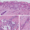
Histology shows orthokeratosis, spongiosis of the epidermis, lymphocytes extravasation is seen in the epidermis, dilated upper-dermis blood vessels with erythrocyte extravasation in all dermis levels (haematoxylin and eosin staining; original magnification 200×). (B) Deep perivascular infiltrate composed mostly of neutrophils with abundant nuclear dust; the inflammatory cells extend to the fat tissue; adnexal structures are unaffected. However, there is a mild inflammatory cell (haematoxylin and eosin staining; original magnification 200×). (C) higher magnification of (A) shows fibrinoid necrosis, neutrophilic nuclear dust (haematoxylin and eosin staining; original magnification 400×)

Similar articles

References

    1. Snow IM. Purpura, urticaria and angioneurotic edema of the hands and feet in a nursing baby. JAMA 1913; 61: 18-9.
    1. Miconi F, Cassiani L, Savarese E, et al. . Targetoid skin lesions in a child: acute hemorrhagic oedema of infancy and its differential diagnosis. Int J Environ Res Public Health 2019; 16: 823. - PMC - PubMed
    1. Jindal SR, Kura MM. Acute hemorrhagic edema of infancy-a rare entity. Indian Dermatol Online J 2013; 4: 106-8. - PMC - PubMed
    1. McDougall C, Ismail SK, Ormerod A. Acute haemorrhagic oedema of infancy. Arch Dis Child 2005; 90: 316. - PMC - PubMed
    1. Carder KR. Hypersensitivity reactions in neonates and infants. Dermatol Ther 2005; 18: 160-75. - PubMed