. 2022 Aug 4:29:643-655.
doi: 10.1016/j.omtn.2022.08.004. eCollection 2022 Sep 13.

Enhancing arginase 2 expression using target site blockers as a strategy to modulate macrophage phenotype

Affiliations

Enhancing arginase 2 expression using target site blockers as a strategy to modulate macrophage phenotype

Chiara De Santi et al. Mol Ther Nucleic Acids. .

Abstract

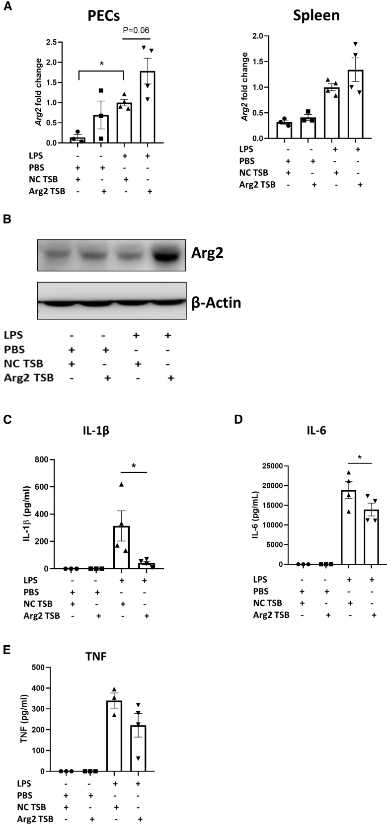
Macrophages are plastic cells playing a crucial role in innate immunity. While fundamental in responding to infections, when persistently maintained in a pro-inflammatory state they can initiate and sustain inflammatory diseases. Therefore, a strategy that reprograms pro-inflammatory macrophages toward an anti-inflammatory phenotype could hold therapeutic potential in that context. We have recently shown that arginase 2 (Arg2), a mitochondrial enzyme involved in arginine metabolism, promotes the resolution of inflammation in macrophages and it is targeted by miR-155. Here, we designed and tested a target site blocker (TSB) that specifically interferes and blocks the interaction between miR-155 and Arg2 mRNA, leading to Arg2 increased expression and activity. In bone marrow-derived macrophages transfected with Arg2 TSB (in the presence or absence of the pro-inflammatory stimulus LPS), we observed an overall shift of the polarization status of macrophages toward an anti-inflammatory phenotype, as shown by significant changes in surface markers (CD80 and CD71), metabolic parameters (mitochondrial oxidative phosphorylation) and cytokines secretion (IL-1β, IL-6, and TNF). Moreover, in an in vivo model of LPS-induced acute inflammation, intraperitoneal administration of Arg2 TSB led to an overall decrease in systemic levels of pro-inflammatory cytokines. Overall, this proof-of-concept strategy represent a promising approach to modulating macrophage phenotype.

Keywords: MT: non-coding RNAs; PLGA; arginase 2; macrophages; miR-155; microRNAs; target site blocker; transfection.


Conflict of interest statement

The authors have no relevant conflicts of interest to declare other than the receipt of funding form the named agencies to carry out the current work.

Figures

None
Graphical abstract
Figure 1
Figure 1
Arg2 TSB effectively blocks miR-155-mediated repression of Arg2 Data are presented as mean ± SEM and were compared by two-way ANOVA (using multiple comparisons test, ∗p ≤ 0.05, ∗∗p < 0.01, ∗∗∗p < 0.001, ∗∗∗∗p < 0.0001). (A) Visual map of the in silico predictions of miR-155 binding to its MRE within Arg2 3′ UTR. (B) Arg2 3′ UTR luciferase activity reported as percent change in relative light units (RLU) in RAW 264.7 cells. Site-directed mutagenesis was applied to pmir_Arg2_wt vector to disrupt the miR-155 MRE. RAW 264.7 cells were co-transfected with wt (first to fourth bars) or miR_155 mutant (fifth to eighth bars) plasmid and TSB and miR mimic (100 ng plasmid, 100 nM TSB, 25 nM mimic, n = 3, in triplicate). Samples co-transfected with WT plasmid, non-targeting control (NC) TSB and NC miR mimic were used as reference and set at 100%. For both WT and miR_155 mutant plasmids, only the comparison between NC and Arg2 TSB (co-transfected with NC or miR-155 mimics) are highlighted in the graph. (C) MiR-155 expression upon stimulation with LPS (100 ng/mL, 24 h) and TSB transfection in primary BMDM (n = 4, in triplicate). LPS-stimulated samples transfected with NC TSB are set as 1. (D) Arg2 TSB effect on endogenous levels of Arg2 mRNA in primary BMDM (n = 4, in triplicate). Unstimulated samples transfected with NC TSB are set as 1. (E) Arg2 TSB effect on endogenous levels of Arg2 protein in primary BMDM (n = 3, in single). LPS-stimulated samples transfected with NC TSB are set as 1. (F) Arginase activity assay in primary BMDM upon transfection of Arg2 TSB (n = 3, in triplicate). Urea was measured as a byproduct of arginase activity and fold arginase activity was measured by setting LPS-stimulated samples transfected with NC TSB as 1.
Figure 2
Figure 2
Arg2 TSB modulates surface markers and metabolic parameters in primary BMDM (A–D) Surface markers expression. Pro-inflammatory markers CD80 (A) and CD86 (B) and anti-inflammatory markers CD71 (C) and CD206 (D) surface levels in Arg2 vs NC TSB transfected BMDM (in presence and absence of LPS) were compared by flow cytometry (n = 3, in single). Histograms depict the results obtained in one representative experiment. The bar graphs represent the means of mean fluorescence intensity ± SEM of positive cells in three biological replicates. Data were compared by one-way ANOVA (Sidak’s multiple comparisons test, ∗p ≤ 0.05, ∗∗p < 0.01, ∗∗∗∗p < 0.0001). (E) Representative Seahorse metabolic flux trace of BMDM transfected with Arg2 vs NC TSB in absence (left) or presence (right) of LPS (10 ng/mL) (n = 5, 4–7 technical replicates). (F) Quantitative oxidative parameters changes in BMDM transfected with Arg2 vs NC TSB in absence or presence of LPS (n = 5, 4–7 technical replicates). Data were compared by two-way ANOVA (Tukey’s multiple comparisons test, ∗∗p < 0.01, ∗∗∗p < 0.001, ∗∗∗∗p < 0.0001).
Figure 3
Figure 3
Arg2 TSB encapsulated in biocompatible PLGA NPs decreases pro-inflammatory cytokines secretion in primary BMDM (A) Pro-inflammatory cytokines secretion by BMDM upon transfection of Arg2 vs NC TSB using the classical transfection reagent lipofectamine 3000 in presence or absence of LPS (n = 3, in triplicate). Samples transfected with NC TSB were used as reference and set at 100%. (B, C) Physicochemical characterization of empty, NC TSB, and Arg2 TSB PLGA NPs using the Zetasizer system for measuring (B) size (nm) on the left y axis (bar charts), poly-dispersity index (PDI) on the right y axis (topmost dots) and (C) surface charge (Z-potential, mV). The values represented are the mean averages ± SEM of at least five independent NPs preparation. (D) Representative images of PLGA NPs stained with uranyl acetate replacement stain using transmission electron microscopy (TEM). (E, F) Effect of Arg2 TSB transfection (with lipofectamine 3000 transfection reagent, LF, second and third bars) and PLGA-TSBs NPs (fourth to sixth bars) on primary BMDM (E) viability (3-(4,5-dimethylthiazol-2-yl)-5-(3-carboxymethoxyphenyl)-2-(4-sulfophenyl)-2H-tetrazolium = MTS assay, n = 3, in triplicate) and (F) toxicity (lactate dehydrogenase = LDH assay, n = 3, in triplicate). (G–I) (G) IL-1β, (H) IL-6, and (I) TNF cytokines secretion by BMDM upon transfection of Arg2 vs NC TSB encapsulated into PLGA NPs in presence or absence of LPS (n = 3, in triplicate). Samples transfected with NC TSB-PLGA NPs were used as a reference and set at 100%.
Figure 4
Figure 4
Arg2 TSB delivery decreases pro-inflammatory cytokines secretion in an LPS in vivo model WT C57Bl/6J mice were given i.p. injection of Arg2 vs NC TSB for 24 h followed by LPS at 5 mg/kg (or PBS control) for 8 h. (A) Arg2 mRNA expression in PECs (left) and spleen (right). Data are presented as mean ± SEM and were compared by one-way ANOVA where mice injected with NC TSB and LPS were set at 1. (B) Arg2 and β-actin in spleen. Blot representative of seven mice from each TSB injection (three of which were then injected with PBS and four with LPS for 8 h). (C) Pro-inflammatory cytokines levels in serum in the LPS model of acute inflammation. Data are presented as mean ± SEM and were compared by one-way ANOVA (Sidak’s multiple comparison test, ∗p < 0.05).

Similar articles

Cited by

References

    1. Ross E.A., Devitt A., Johnson J.R. Macrophages: the good, the bad, and the gluttony. Front. Immunol. 2021;12:708186. doi: 10.3389/fimmu.2021.708186. - DOI - PMC - PubMed
    1. Kałużna A., Olczyk P., Komosińska-Vassev K. The role of innate and adaptive immune cells in the pathogenesis and development of the inflammatory response in ulcerative colitis. J. Clin. Med. 2022;11:400. doi: 10.3390/jcm11020400. - DOI - PMC - PubMed
    1. Jang S., Kwon E.J., Lee J.J. Rheumatoid arthritis: pathogenic roles of diverse immune cells. Int. J. Mol. Sci. 2022;23:905. doi: 10.3390/ijms23020905. - DOI - PMC - PubMed
    1. Nally F.K., De Santi C., McCoy C.E. Nanomodulation of macrophages in multiple sclerosis. Cells. 2019;8 doi: 10.3390/cells8060543. - DOI - PMC - PubMed
    1. Oishi Y., Manabe I. Macrophages in inflammation, repair and regeneration. Int. Immunol. 2018;30:511–528. doi: 10.1093/intimm/dxy054. - DOI - PubMed