. 2022 Aug 4:29:625-642.
doi: 10.1016/j.omtn.2022.07.027. eCollection 2022 Sep 13.

Identification and characterization of a MAPT-targeting locked nucleic acid antisense oligonucleotide therapeutic for tauopathies

Affiliations

Identification and characterization of a MAPT-targeting locked nucleic acid antisense oligonucleotide therapeutic for tauopathies

Amy Easton et al. Mol Ther Nucleic Acids. .

Abstract

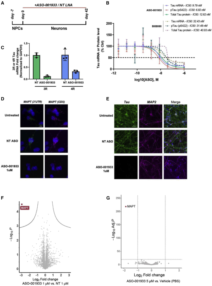
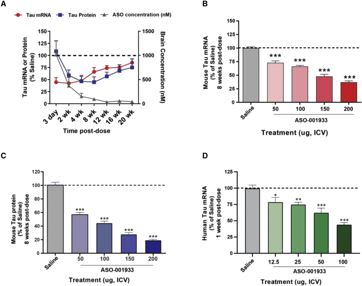
Tau is a microtubule-associated protein (MAPT, tau) implicated in the pathogenesis of tauopathies, a spectrum of neurodegenerative disorders characterized by accumulation of hyperphosphorylated, aggregated tau. Because tau pathology can be distinct across diseases, a pragmatic therapeutic approach may be to intervene at the level of the tau transcript, as it makes no assumptions to mechanisms of tau toxicity. Here we performed a large library screen of locked-nucleic-acid (LNA)-modified antisense oligonucleotides (ASOs), where careful tiling of the MAPT locus resulted in the identification of hot spots for activity in the 3' UTR. Further modifications to the LNA design resulted in the generation of ASO-001933, which selectively and potently reduces tau in primary cultures from hTau mice, monkey, and human neurons. ASO-001933 was well tolerated and produced a robust, long-lasting reduction in tau protein in both mouse and cynomolgus monkey brain. In monkey, tau protein reduction was maintained in brain for 20 weeks post injection and corresponded with tau protein reduction in the cerebrospinal fluid (CSF). Our results demonstrate that LNA-ASOs exhibit excellent drug-like properties and sustained efficacy likely translating to infrequent, intrathecal dosing in patients. These data further support the development of LNA-ASOs against tau for the treatment of tauopathies.

Keywords: MAPT; MT: oligonucleotides: therapies and applications; antisense oligonucleotides; cynomolgus monkey; intrathecal; locked nucleic acids; neurodegeneration; tau; tauopathies.


Conflict of interest statement

C.M.K., J.K.L., R.S., Y.B., D.D., S.E.M., and R.E.O. are employees of BMS and own stock or restricted stock units in BMS. A.E., Y. Li, Y. Lu, J.E. Meredith, J.E. Macor, M.W., V.W., K.J., M.G., J.M.B., L.H., A.F., J.P., M.B., A.B., J.E.M., C.F.A., and A.M.C. were employees of BMS when the work described was carried out. R.E.O., A.M.C., P.H.H., A.M.H., J.M.B., M.L.J., and S.E.M. are co-inventors on US Patent 10,799,523 and US patent applications US 2016/0237427, US 2018/0161356 and US 2019/0383797; and PCT patent application 2016/126995. P.H.H., R.E.O., A.M.H., and M.L.J. are co-inventors on US patent application US 2018/0023081.

Figures

None
Graphical abstract
Figure 1
Figure 1
Initial screen identified parental MAPT-targeting sequence ASO-000013 (A and B) In vitro ASO-tiling and toxicity screen using hTau mouse primary neuronal cultures. Quantitative analysis of Tau protein levels after ASO treatments. ASO-targeting sites along MAPT transcript are shown on the x axis (A). Calcium oscillations (CaOsc) and β-ΙΙΙ tubulin levels were evaluated for all ASOs used in tiling experiment (B). Each spot represents an ASO. Forty-seven ASOs (shown in green) showed low in vitro toxicity (tubulin >70% UTC and CaOsc >75% UTC) and good target knockdown efficiency (>80% UTC, B). (C) Thirteen out of 47 ASOs were selected for further evaluation using hTau mice. Assessment of in vivo acute tolerability and relative MAPT expression for each selected ASO. Each spot represents an ASO. ASO (shown in green) was selected as a parental MAPT-targeting sequence ASO-000013. (D) Alternative splicing of the MAPT gene generates six tau isoforms, named after the number of N-terminal repeats (0 N, 1 N, and 2 N) and C-terminal microtubule-binding domain repeats (3R and 4R). The first two exons (−1, 1) encode the 5′ untranslated region (UTR) and exon 14 encodes the 3′ UTR. The exons 4a, 6, and 8 are only part of the mature tau transcript in the peripheral nervous system. The target RNA sequence of ASO-000013 in exon 14 of tau mRNA is highlighted in green. The surrounding sequence in human, Cyno monkey, mouse, and rat show high conservation across different species. Nucleotides in red differ from the human sequence.
Figure 2
Figure 2
LNA pattern modifications altered ASO potency and tolerability both in vitro and in vivo (A) IC50 (protein) and tolerability of hTau mouse primary neuronal cultures upon ASO treatments. Corresponding LNA pattern modifications are displayed along the x axis (L, LNA; D, DNA). ASOs with good tolerability (tubulin >70% UTC and CaOsc > 75%UTC), activity, and selectivity were selected for in vivo study. (B) Dose-dependent reduction of MAPT expression at protein level upon ASO-001933 treatment. hTau mouse primary neurons were treated with ASO-001933 at indicated concentrations. Tau protein levels were measured by high-content imaging (n = 15). (C) Assessment of in vivo acute tolerability and relative MAPT expression for each selected ASO. Each spot represents an ASO. ASO-001933 is shown in red.
Figure 3
Figure 3
ASO-001933 is a highly potent and selective ASO targeting MAPT on human neurons (A) Schematic diagram of ASO treatment schedule in hESCs for (B)–(F). (B) Dose-dependent reduction of MAPT expression at RNA and protein level upon ASO-001933 or BIIB080 treatment. hESC-derived neurons were treated with ASO at indicated concentrations. RNA and protein levels were assessed by qRT-PCR and AlphaLISA respectively. Absolute IC50 values are reported in the figure (n = 3 /treatment, mean +/- SEM). (C) Quantification of 3R and 4R Tau mRNA by qPCR after ASO treatment at 1 μM (n = 3 /treatment, mean +/- SEM). (D and E) Representative images of RNAscope ISH and ICC using probes against MAPT CDS, MAPT 3′ UTR (D) and antibody against Tau (Tau HT7) (E). (F) Volcano plot illustrating differentially regulated proteins from proteomics analysis between ASO-001933-treated cells versus NT ASO-treated neurons (n = 3). MAPT is the only significantly regulated protein (Log2 FC = −2.94, adjusted Log10 p value = −4.54). (G) Evaluation of off-target effects of ASO-001933 in hiPSC-derived neurons. Volcano plot showing differentially expressed genes (Log2FC > 1 or Log2FC< −1 and −Log10 adjusted p >20) from RNA-seq analysis after 72 h of treatment (n = 3).
Figure 4
Figure 4
Single dose of ASO-001933 shows a robust and sustained reduction of mouse and human Tau in vivo (A) ASO-001933 (100 μg ICV) time course for mouse tau mRNA (red circles) and tau protein (blue squares) in C57Bl/6J mice. ASO-001933 concentrations (gray triangles) in brain over the time course. (B) mTau mRNA is decreased by 70% 8 weeks after a single injection of 200 μg (n = 10–20/group, mean ± SEM). (C) ASO-001933 reduces mTau protein by 80% 8 weeks after a single injection (n = 9–19, mean ± SEM). (D) ASO-001933 reduces hTau mRNA by 1 week post injection (n = 10/group, mean ± SEM). Statistical analysis: one-way ANOVA with Dunnett’s post hoc, ∗p < 0.05, ∗∗p < 0.01, ∗∗∗p < 0.0001.
Figure 5
Figure 5
Broad ASO-001933 brain uptake and tau transcript lowering in NHP brains after IT delivery ASO-001933 uptake in the frontal cortex of NHPs dosed with ASO-001933 at 1 × 8 mg (A), 2 × 8 mg (B), 2 × 16 mg (C), and vehicle (D), respectively. ASO-001933 is visualized with a fluorescently labeled sense probe. Pseudocolor green indicates ASO-001933 uptake. (E and F) Tau mRNA expression in the forebrain coronal section from the NHP dosed with vehicle or ASO-001933 at 2 × 16 mg. Tau mRNA expression is visualized with a 35S-labeled antisense probe to Tau mRNA. Pseudocolor yellow/red indicates tau transcript expression. (G and H) Tau mRNA expression in the ventral region of the midbrain (upper) and pons (lower) of the NHP dosed with vehicle or ASO-001933 at 2 × 16 mg. Tau mRNA is visualized with a 35S-labeled antisense probe to Tau mRNA. Pseudocolor yellow indicates tau transcript expression. SN, substantia nigra; PN, pontine nuclei. n = 1 or 2 monkeys/treatment group.
Figure 6
Figure 6
ASO-001933 has a long duration of action on tau in both brain and CSF in NHP brains after IT delivery ASO-001933 (2 × 8 mg) produces robust mRNA and protein reduction in frontal cortex (A) and hippocampus (B). Brain tau protein lowering (average of the following brain regions: ventral midbrain, ventral pons, medulla, cerebellum, parietal cortex, frontal cortex, and hippocampus) predicted CSF tau protein lowering (C). Vertical dashed red lines indicate time of first and second 8-mg dose, respectively. Tau lowering was detected in all brain regions assessed with highest knockdown in frontal cortex and less in ventral midbrain (D). For CSF tau, n = 2 control and n = 6 ASO-treated monkeys. For brain tau, n = 3 vehicle-treated and n = 1 ASO-treated monkey at 6, 18, and 22 weeks post first dose, n = 2 ASO treated at 10 and 14 weeks post dose. UTC, untreated control. Data are represented as the group mean ± SEM.
Figure 7
Figure 7
PK/PD modeling of ASO-001933 in mouse (A) Steady state achieved with 200-μg dose once every 16 weeks, and, in NHP (B), steady state is achieved with 8-mg dose once every 20 weeks.

Similar articles

Cited by

References

    1. Weingarten M.D., Lockwood A.H., Hwo S.Y., Kirschner M.W. A protein factor essential for microtubule assembly. Proc. Natl. Acad. Sci. USA. 1975;72:1858–1862. - PMC - PubMed
    1. Chang C.W., Shao E., Mucke L. Tau: enabler of diverse brain disorders and target of rapidly evolving therapeutic strategies. Science. 2021;371:eabb8255. - PMC - PubMed
    1. Binder L.I., Frankfurter A., Rebhun L.I. The distribution of Tau in the mammalian central nervous system. J. Cell Biol. 1985;101:1371–1378. - PMC - PubMed
    1. LoPresti P., Szuchet S., Papasozomenos S.C., Zinkowski R.P., Binder L.I. Functional implications for the microtubule-associated protein tau: localization in oligodendrocytes. Proc. Natl. Acad. Sci. USA. 1995;92:10369–10373. - PMC - PubMed
    1. Trushina N.I., Bakota L., Mulkidjanian A.Y., Brandt R. The evolution of Tau phosphorylation and interactions. Front. Aging Neurosci. 2019;11:256. - PMC - PubMed